
My project is about hyoid bone fractures and covers multiple objectives, such as classification of fractures, the bone shape, location of the bone, and functions of the bone.
The hyoid bone is a small, U-shaped solitary bone located in the anterior neck at the base of the mandible and posteriorly at the fourth cervical vertebra. It is a part of the axial skeleton. The hyoid is involved in all of the oral complex functions like tongue movement, mastication, swallowing, regurgitation prevention, and breathing. The hyoid keeps the head in the correct position because of the complicated connection it makes between the jawbone and the spine (AlJulaih et al., 2023).
Hyoid bone fractures are uncommon and usually associated with strangulation. Sharp anterior neck pain that becomes worse while talking, eating, coughing, or swallowing are typical symptoms of the fracture (Porr et al., 2012). Also, patients may experience pain with head rotation, swelling in the neck, and hoarseness. Due to its proximity to the surrounding structures, fractures of the hyoid bone are often combined with fractures of the mandible, cervical spine, larynx, and neck (Alwasiyah et al., 2023).
There are many ways to diagnose the fracture, such as physical examination, cervical radiography. CT scan, laryngoscopy, nasendoscopy or surgical inspection (Porr et al., 2012). However, it may not be possible to detect hyoid fractures immediately because of the more urgent nature of these injuries. In such cases, failure to diagnose or delay of diagnosis may have a negative impact on mortality.
The severity of symptoms is the primary factor determining treatment for hyoid bone fractures. conservative treatment is normally sufficient for closure or asymptomatic fractures, and usually no surgical intervention is required. Soft neck rest fixation, and liquid diets can be used while healing from the fracture. It is also recommended for patients to be observed for 48-72 hours to avoid any complications such as hemoptysis, edema, ecchymosis, and spasm (Alwasiyah et al., 2023).
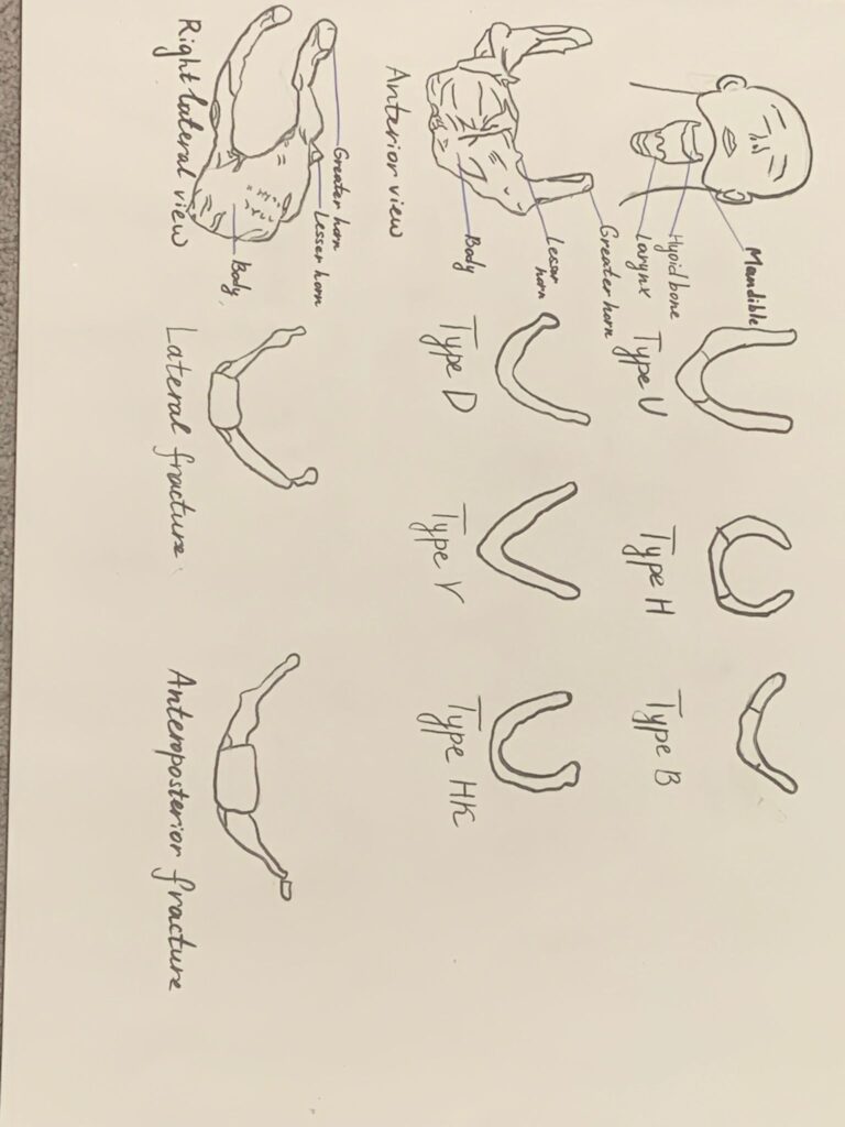
In my project, I illustrated the anatomy of the hyoid bone (on the left) as well as the types of hyoid bones (on the top right) and types of fractures (on the bottom right). The hyoid bone I drew shows the main body the front portion of the bone that is usually smaller in females), greater horns (make up the sides that visibly remind “U”), and lesser horns (that’s where stylohyoid ligament attaches). The greater and the lesser horns are connected by fibrous tissue or a true joint to the body of the hyoid (AlJulaih et al., 2023).
I included two types of fractures: lateral fracture and anteroposterior compression fracture. In the first case, we can see how the bone fracture is displayed inwards. In the second case, it is outwards.
Work Cited
AlJulaih, G. H., & Menezes, R. G. (2019, April 5). Anatomy, Head and Neck, Hyoid Bone. Nih.gov; StatPearls Publishing. https://www.ncbi.nlm.nih.gov/books/NBK539726/
Alwasiyah, B. M., Aljehani, Z. H., Farag, N. H., Alwasiyah, B., Aljehani, Z., & Farag, N. H. (2023). A Rare Isolated Hyoid Bone Fracture: A Case Report and Review of the Literature. Cureus, 15(4). https://doi.org/10.7759/cureus.37165
Porr, J., Laframboise, M., & Kazemi, M. (2012). Traumatic hyoid bone fracture – a case report and review of the literature. The Journal of the Canadian Chiropractic Association, 56(4), 269–274. https://www.ncbi.nlm.nih.gov/pmc/articles/PMC3501913/
This steam project was describing the Hyoid bone and some of the various types of fractures it is susceptible to. The hyoid bone is uncommonly fractured, but when it is, it’s done so by trauma sustained by strangulation. The illustrations reflected well on the various shapes, fractures, and overall location of the hyoid bone. The description of the hyoid bones function, and it’s make up of the oral complex functions were well described. I learned that when the hyoid bone is fractured, it can be difficult to diagnose due to the fact that this bone is usually damaged along with structures such as the mandibles, which is a more urgent trauma to address, thus meaning the fracture of the hyoid bone could result in delayed diagnosis. As stated in the report, this delay could result in a negative impact on mortality.
The art portion of this project was very well executed. Looking at the various illustrations of the hyoid bone really demonstrates just how different each type of bone shape could be, along with the description in the report stating that those shapes could be smaller (in the case of a female), or larger (male). The illustration showing where this bone is located in comparison to the human anatomy was especially interesting and helpful in understanding just where it’s located within the body. Overall this project was really interesting and informative and the student demonstrated a wonderful understanding of the objective. Reflecting that understanding through this project, and thus helped me grasp and understand the objective better.