
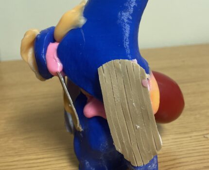
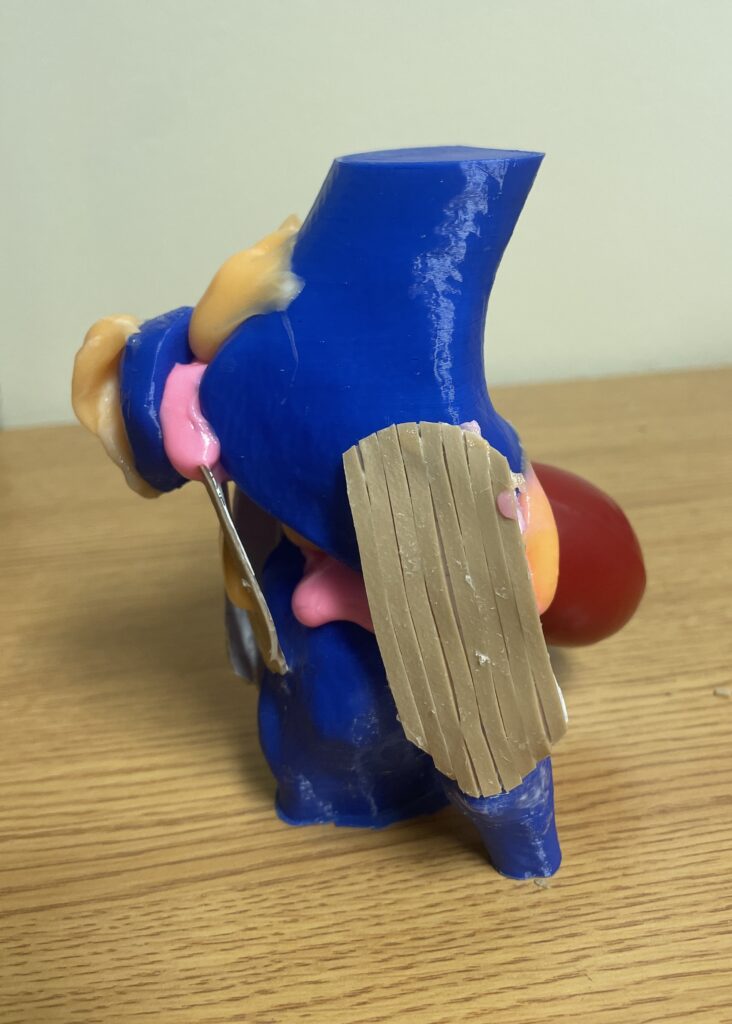
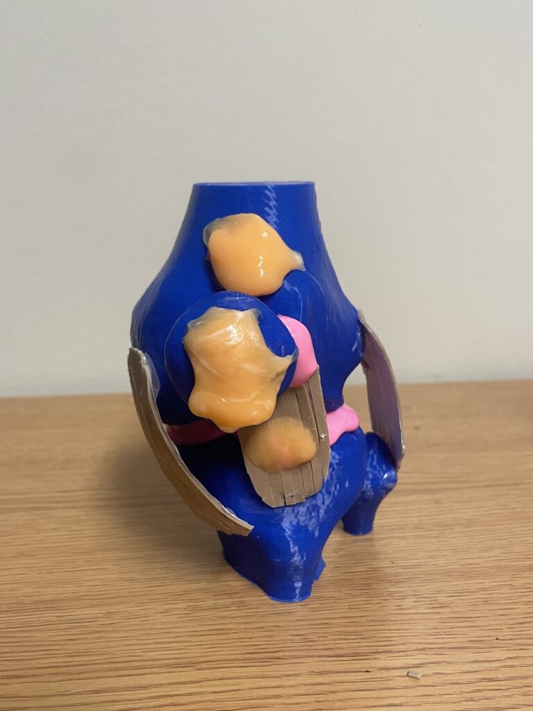
My project was on Baker’s Cyst, a fluid filled cyst that forms from a bursa at the back of the knee joint. A bursa’s function is to reduce friction when moving the joint, but when excessively filled, it is related to many problems like intra-articular knee disorders. This includes the synovial joint and relates the basic structure of a knee joint. The rubber bands are meant to be ligaments, pink colored putty is the cartilage, orange colored putty is the bursas, and the red balloon is the Baker’s cyst. The material is meant to be similar to function of the structure and this model only includes very basic identifying bone, ligament, and bursas structures. The cleanliness of the model was difficult because the putty was very hard to work with on the filament of the model, so it kept moving as I molded it. Hope you learn something! (Photo orientation order: left side with Baker’s cyst, left side without Baker’s cyst, right side with Baker’s cyst, front, back without Baker’s cyst).


The essay written by Michelle Ramirez clearly demonstrates an in-depth dive into the structure of the knee joint. Her essay explains why the knee joint is a synovial joint. She also explains that joints are characterized by their mobility and explains the difference between synarthrosis, amphiarthrosis, and diarthrosis with a good conclusion that synovial joints are uniaxial diarthrosis. She takes the time to describe the structures of her 3D printed model but also to touch on the structures not included. I enjoyed that she switched colors and materials when demonstrating the different structures of the knee as it makes it much easier to identify each part of her model. The creative use of the rubber bands to represent ligaments because they are made of dense fibrous connective tissue proper is clever and appealing to the eye when examining her model.
I was not aware that the knee joint contained so many bursae, but Michelle breaks it down in her essay to explain that the knee has prepatellar, infrapatellar, and suprapatellar bursa. Great use of Visible body as a resource for her project. I would have liked a bit more information about baker’s cyst itself, the symptoms and presentation of the baker’s cyst, and what the underlying conditions are that can cause a baker’s cyst. Information on where and why the excess fluid of the knee creates the cyst could be helpful to other readers in the future. All and all this presentation does a excellent job at identify the structures of the knee, staying true to the objectives learned in class, and indicates a good understanding of multiple units touched on this semester. Really well done!