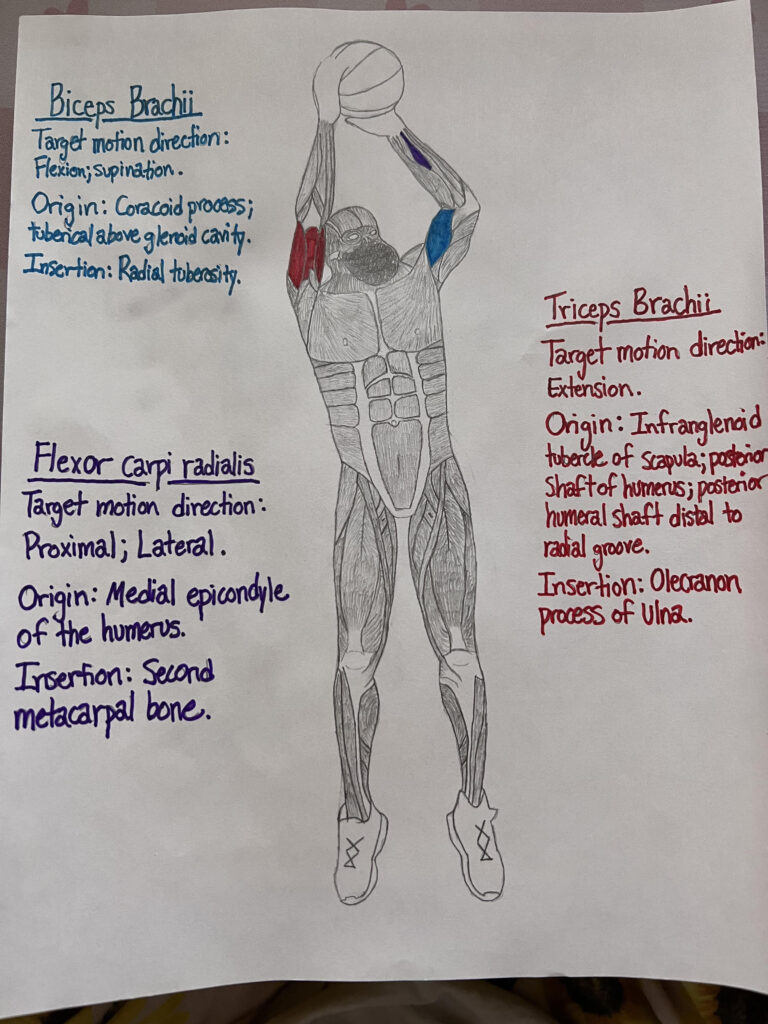
This is my STEAM PROJECT medium. It is a basketball player in the midst of a jump shot. My course objective is to identify the main muscles that are used during a jump shot and to type a 500+ essay about describing how a muscle contraction is induced, which to my knowledge, will be posted in Canvas:) Thank you!
2 Comments
Joshua’s STEAM project objective is to identify the main muscles that are used during a jump shot. In his visual, he did a very detailed person making a jump shot and identified all the muscles that are involved during this action and put their target motion direction, origin, and insertion. These muscles are the biceps brachii, triceps brachii, and flexor carpi radialis.
Cheyanne Coglon
~ During the action of pulling you up off the earth, you have your lower limb skeletal muscles pushing you up into the air, but what about taking the fatal shot? According to Pakosz, P., (2011). Emg signal analysis of selected muscles during shots and passes in basketball. 2, 12. he presents a table consisting of active muscles during shots and passes. It shows active EMG activity of primarily the triceps/biceps brachii during a jump-shot. Lets examine how these muscles are contracted molecularly. Bagshaw, C. R., (1993). Muscle Contraction, Relating to the beginning of muscle contraction and action potential, states that, the ratio of external to internal positive potassium concentration in a resting nerve is nearly 0.1 and implies that this ion is almost at equilibrium. In contrast, positive sodium is maintained and mended at a ratio of 10 by an active sodium pump. In excitation, the nerve cell membrane becomes locally and temporarily permeable to NA+, which results in its concentration gradient to rapidly deplete, causing the cell’s membrane potential to rise to +40 mV. This relates to how nerves control skeletal muscles and how they partake a big part in the contraction process. The beginning of the muscle contraction process starts off with this action potential, or AP, sent from the nerve heading to the muscle and then arriving at the axon terminal. Geeves, M. A., & Holmes, K. C., (2005). The Molecular Mechanism of Muscle Contraction (71st ed.). explains in their book that the myosin structure has been unraveled in a number of different conformations. Actin appears to be the more passive partner—providing binding sites for cross bridges as its main and top primary function. This is an illustration of how in the next step of muscle contraction, cross bridges are formed by actin and the Myosin head. More from Geeves M., explains that the binding to actin also enables the release of products. Phosphate is first released from the cross bridge followed by Adenosine diphosphate. Nearing the end of the power-stroke, ADP is released, which allows a new Adenosine triphosphate molecule to bind and connect to the myosin. The cross bridge triggers a rapid release from the actin filament and the cycle starts again. Heading back to the main topic of relating to basketball, Miller, S., (1997). 15 International Symposium on Biomechanics in Sports. Contribution of Selected Muscles to Basketball Shooting, records and reports that high traces of EMG activity were located in 4 main muscles. These muscles include the Bicep brachii; Tricep brachii; Flexor carpi radialis; and Extensor carpi radialis. The action potential releases acetylcholine, which is a transmitter substance. In which it depolarizes the muscle cell membrane. Fibre membranes are highly invaginated and carry an electrical signal. That electrical signal is then sent inwards and results in the sarcoplasmic reticulum to become stimulated inside of the adjacent vesicles. This then leads to the release CA2+. Bagshaw, C. R., (1993). This is relating to the last process of muscle contraction. Hydrolyzation of Adenosine triphosphate to Adenosine diphosphate and phosphate, restoring back to the original state and posses the ability to restart the process all back to the beginning. This finalizing the process of muscle contraction and the relation in the main muscles used while taking a jump shot.

Joshua’s STEAM project objective is to identify the main muscles that are used during a jump shot. In his visual, he did a very detailed person making a jump shot and identified all the muscles that are involved during this action and put their target motion direction, origin, and insertion. These muscles are the biceps brachii, triceps brachii, and flexor carpi radialis.
~ During the action of pulling you up off the earth, you have your lower limb skeletal muscles pushing you up into the air, but what about taking the fatal shot? According to Pakosz, P., (2011). Emg signal analysis of selected muscles during shots and passes in basketball. 2, 12. he presents a table consisting of active muscles during shots and passes. It shows active EMG activity of primarily the triceps/biceps brachii during a jump-shot. Lets examine how these muscles are contracted molecularly. Bagshaw, C. R., (1993). Muscle Contraction, Relating to the beginning of muscle contraction and action potential, states that, the ratio of external to internal positive potassium concentration in a resting nerve is nearly 0.1 and implies that this ion is almost at equilibrium. In contrast, positive sodium is maintained and mended at a ratio of 10 by an active sodium pump. In excitation, the nerve cell membrane becomes locally and temporarily permeable to NA+, which results in its concentration gradient to rapidly deplete, causing the cell’s membrane potential to rise to +40 mV. This relates to how nerves control skeletal muscles and how they partake a big part in the contraction process. The beginning of the muscle contraction process starts off with this action potential, or AP, sent from the nerve heading to the muscle and then arriving at the axon terminal. Geeves, M. A., & Holmes, K. C., (2005). The Molecular Mechanism of Muscle Contraction (71st ed.). explains in their book that the myosin structure has been unraveled in a number of different conformations. Actin appears to be the more passive partner—providing binding sites for cross bridges as its main and top primary function. This is an illustration of how in the next step of muscle contraction, cross bridges are formed by actin and the Myosin head. More from Geeves M., explains that the binding to actin also enables the release of products. Phosphate is first released from the cross bridge followed by Adenosine diphosphate. Nearing the end of the power-stroke, ADP is released, which allows a new Adenosine triphosphate molecule to bind and connect to the myosin. The cross bridge triggers a rapid release from the actin filament and the cycle starts again. Heading back to the main topic of relating to basketball, Miller, S., (1997). 15 International Symposium on Biomechanics in Sports. Contribution of Selected Muscles to Basketball Shooting, records and reports that high traces of EMG activity were located in 4 main muscles. These muscles include the Bicep brachii; Tricep brachii; Flexor carpi radialis; and Extensor carpi radialis. The action potential releases acetylcholine, which is a transmitter substance. In which it depolarizes the muscle cell membrane. Fibre membranes are highly invaginated and carry an electrical signal. That electrical signal is then sent inwards and results in the sarcoplasmic reticulum to become stimulated inside of the adjacent vesicles. This then leads to the release CA2+. Bagshaw, C. R., (1993). This is relating to the last process of muscle contraction. Hydrolyzation of Adenosine triphosphate to Adenosine diphosphate and phosphate, restoring back to the original state and posses the ability to restart the process all back to the beginning. This finalizing the process of muscle contraction and the relation in the main muscles used while taking a jump shot.
My Medium Link 🙂
https://humanap.community.uaf.edu/2023/11/21/9125/Links to an external site.
Pakosz, P., (2011). Emg signal analysis of selected muscles during shots and passes in basketball. 2, 12 https://www.researchgate.net/profile/Pawel-Pakosz-2/publication/283150472_EMG_SIGNAL_ANALYSIS_OF_SELECTED_MUSCLES_DURING_SHOTS_AND_PASSES_IN_BASKETBALL/links/562cc92c08aef25a24430130/EMG-SIGNAL-ANALYSIS-OF-SELECTED-MUSCLES-DURING-SHOTS-AND-PASSES-IN-BASKETBALL.pdf
Bagshaw, C. R., (1993). Muscle Contraction,
https://books.google.com/books?hl=en&lr=&id=cEDryw-RXUEC&oi=fnd&pg=PR9&dq=steps+of+muscle+contraction&ots=hIDO-t5Hlx&sig=2vUT05Lqsh3Lv_xrdWE5D6dH7uI#v=onepage&q=steps%20of%20muscle%20contraction&f=falseLinks to an external site.
Geeves, M. A., & Holmes, K. C., (2005). The Molecular Mechanism of Muscle Contraction (71st ed.). https://doi.org/10.1016/S0065-3233(04)71005-0Links to an external site.
Miller, S., (1997). 15 International Symposium on Biomechanics in Sports. Contribution of Selected Muscles to Basketball Shooting, https://ojs.ub.uni-konstanz.de/cpa/article/view/3657