
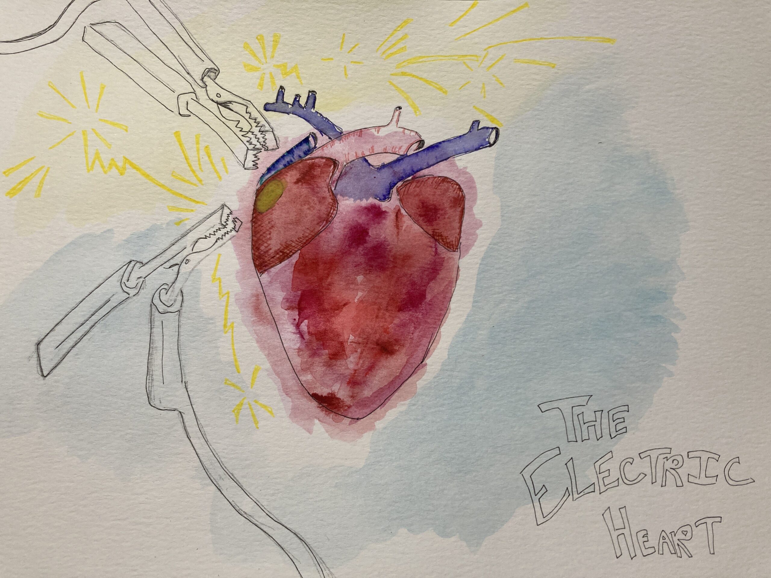
How does a heart beat? What forces work together so that, even as you are reading this article, your heart squeezes itself at least every second? For my STEAM project I created a colorful image of a human heart by using watercolor and colored pens. Attached to the sinoatrial node are jumper cables, illustrating that the electrical signals in the heart are generated at the SA node. It could be interpreted as the SA node receiving a signal from outside the heart, but this is not what truly occurs in our body. In other words, the jumper cables are used to show the start of an electric impulse, not the conduction of an impulse from an outside system.
More specifically, the heart is full of cardiac muscle that is myogenic, which means it can generate a contraction independently of impulses from the central nervous system. This is due to two kinds of cardiac muscle cells in the heart. The heart contains myocardial contractile cells and conducting cells, with contractile cells totalling about 99% of the heart’s composition, with conducting cells making up the remaining 1% (Biga et al). The conducting cells are considered to be autorhythmic, meaning they create and send the electric impulses throughout their conduction system.
The SA node is the beginning of this conduction network, and is known as the pacemaker of the heart. The SA node is located in the atrial wall of the right atrium. Its impulse is sent down to atrial contractile cells and the atrioventricular node, which depolarizes after a slight delay (Goldreyer et al). This delay is important to heart function because it allows the atrial contractile cells to fully contract, sending the blood they contain into the ventricles. Connective tissue in the heart blocks the impulse from reaching the ventricles along any route that is not the AV node (Biga et al). With this funneling, the impulse travels past the AV node at a speed of about 0.12 meters per second, through the atrioventricular bundle, and splits down the right and left bundle branches (Sher et al). These branches wrap around the ventricles and end with the subendocardial conducting network, also known as purkinje fibers. The purkinje fibers are conductive cells that spread the impulse to the contractile cells of the ventricles.
The total time from the generation of the impulse in the SA node, to the depolarization and contraction of the ventricles is about 225 milliseconds (Biga et al). This is how our heart beats, by generating an internal electrical impulse, and contracting in the fraction of a second.
Biga, L. M., Dawson, S., Harwell, A., Hopkins, R., Kaufmann, J., LeMaster, M., Matern, P., Morrison-Graham, K., Quick, D., & Runyeon, J. (2019, September 26). 19.2 cardiac muscle and electrical activity. Anatomy Physiology. Retrieved April 20, 2023, from https://open.oregonstate.education/aandp/chapter/19-2-cardiac-muscle-and-electrical-activity/
Goldreyer, B., & Damato, A. (1971, May 1). The Essential Role of atrioventricular conduction delay in the … Circulation. Retrieved April 21, 2023, from https://www.ahajournals.org/doi/10.1161/01.CIR.43.5.679 Sher, A. M., Rodriguez, M., Liikane, J., & Young, A. C. (1959, January 1). The mechanism of atrioventricular conduction – circulation research. Circulation. Retrieved April 21, 2023, from https://www.ahajournals.org/doi/10.1161/01.RES.7.1.54
Good job on your drawing! The heart is such a powerful and intricate organ. When someone asks me to think of a strong muscle in the body, I tend to overlook the heart. Like you said “the heart is full of cardiac muscle”. As for electrical impulses, I often think of the brain waves and not the heart. As mentioned above “The heart contains myocardial contractile cells and conducting cells”. We know that we can not live without a functioning heart, but rarely think of how it works. The sinoatrial (SA) node and atrioventricular (AV) node are tricky for me to remember. But thanks to your cable jumper illustration, I will be able to remember that SA node “jump starts” the heart. SA node is considered the pacemaker of the heart because its electrical signals cause the atria heart to contract (pump). Then it caused a cascade effect to the AV node causing them to pump. Such a perfect illustration!