
The path of our blood flow through the heart and the circulatory system is amazing. Our deoxygenated blood from the body enters the right side of the heart from the Superior and Inferior Vena Cava. Our oxygenated-rich blood is delivered from the lungs back to the Left side of the heart to be distributed to the rest of the body and its vital organs, tissues, and extremities within the circulatory system and this all happens with each beat of our heart. What makes this so awe-inspiring is that this entire journey takes place about every 60 seconds (Cleveland Clinic, 2022).
At the center of the circulatory system, there is a great muscular pump called the human heart. This pump helps us to function minute-by-minute, hour-by-hour, and day-by-day basis, each day of our lives. The heart is a strong muscle that is almost the size of a person’s fist (Cleveland Clinic), 2022. It is a muscular organ located in the middle of the chest and sits behind the sternum tilted slightly to the left (Rehman, I., & Rehman, A. 2022). The heart beats about 60-100 times per minute and about 100,000 times per day. Every sixty seconds it processes and circulates about 5 liters of blood. What’s even crazier, is that turns out to equal about 2,000 gallons of blood which are pumped through the heart daily; enough to fill a swimming pool that is approximately 8 by 10 ft in size (Cleveland Clinic, 2022).
The ability of the blood to travel through our circulatory systems is very important. Our bodies require oxygenated blood from head to toe to function and live. The right side of our heart receives deoxygenated blood that has just traveled from our body and extremities and the left side of the heart receives oxygenated blood that is returned to it from our lungs (Cleveland Clinic, 2022).
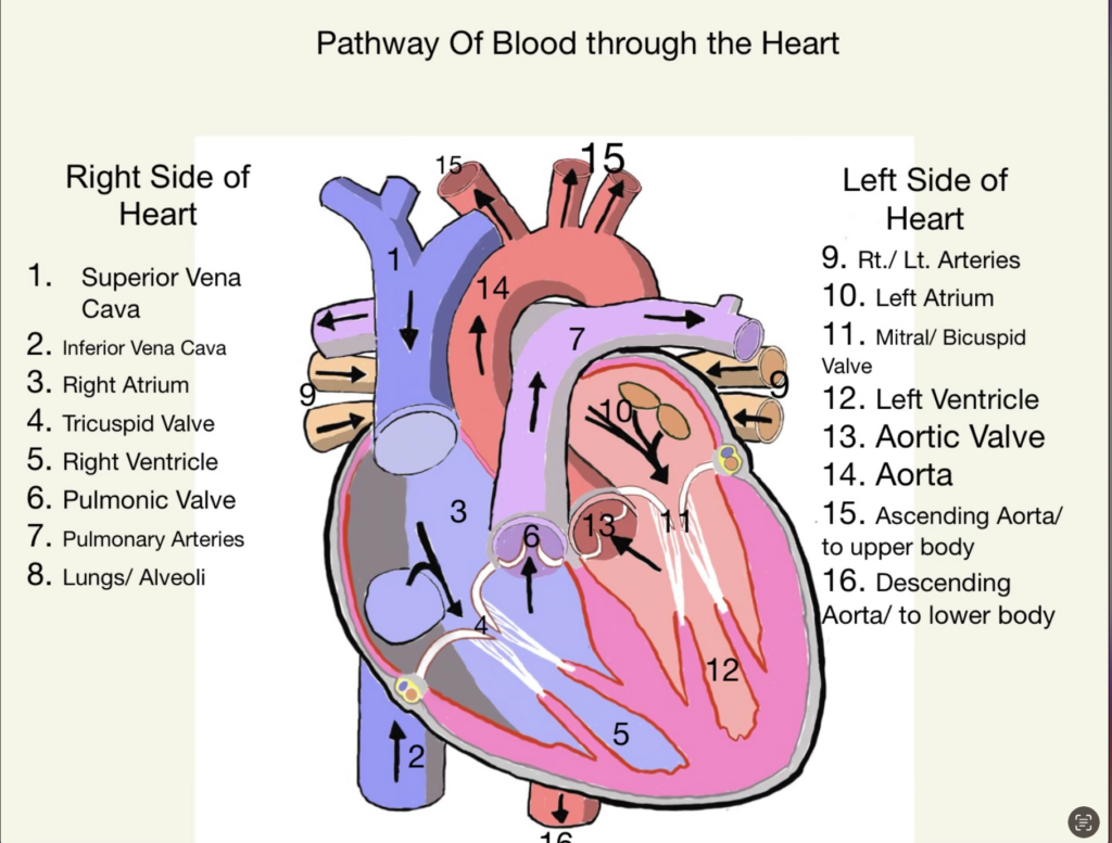
The heart is made up of 4 Chambers. The two upper chambers are called the right and left atria, while the two lower chambers are called the right and left ventricles. The right atrium and ventricle together are often called the Right Heart, and the left atrium and the left ventricle together functionally form the Left Heart. These chambers are divided into the right and left halves. These halves are separated by a muscular wall called a septum (Rehman, I., & Rehman, A. 2022).
The heart receives deoxygenated blood from the extremities of both our lower and upper body. This blood enters a large vein that is divided into two parts. The Superior vena cava receives deoxygenated blood from the upper body, or the regions above our diaphragm. The blood also enters the Inferior vena cava from the lower regions of the body below the diaphragm. This blood enters the top right chamber of the heart called the right atrium. It is then pumped through a valve that separates the upper and lower right chambers called the Tricuspid valve. Once the blood is delivered, this valve closes ensuring that the blood continues to flow in the right direction and this allows it to be pumped into the bottom right chamber of the heart called the Rt. Ventricle. As the Right Ventricle contracts, the tricuspid valve closes and this opens the pulmonary semi-lunar valve. This allows the blood to travel up through the pulmonary trunk and artery (Cleveland Clinic, 2022). What’s fascinating is that the pulmonary artery which is carrying this oxygen-poor blood is blue while most of our other arteries are red. Nonetheless, it’s still an artery because it’s taking blood away from the heart.
The second half of the journey for this deoxygenated blood begins as it enters the lungs via the Right and Left pulmonary arteries. This blood then experiences the process of diffusion. The CO2 in the blood of our pulmonary artery crosses over into the alveoli of our lungs which will later be expired as we breathe out. The O2 in the alveoli of the lungs crosses over into the blood to re-oxygenate it. This blood now continues the second half of its journey via the Right and Left Pulmonary veins. These veins return the oxygenated blood back to the heart and deliver it to the Left Atrium. Once here, the blood is pushed with great force through the Mitral or Bicuspid valve, and then into the Left Ventricle or last chamber of the heart (Sanchez, F., Bordoni, B., 2022). As the Left ventricle contracts, this forces the Aortic Semilunar valve to open and this blood is pushed up through this valve and into the Aorta, which is the largest artery in the body. Next, it travels with great force and speed to deliver oxygen to the vital organs and extremities of both the upper and lower body via the ascending and descending aorta. From here, the blood picks up the oxygen-poor blood that is full of CO2 and delivers it back to the Inferior and Superior Vena Cava to start the process all over again (Rehman, I., & Rehman, A., 2022).
References
Professional, M. (2022, April 29). Blood flow through the heart: Pathways and circulation. Cleveland Clinic. Retrieved April 20, 2023, https://my.clevelandclinic.org/health/articles/17060-how-does-the-blood-flow-thro5ugh-your-heart
Rehman, I., & Rehman, A. (2022, October 19). Anatomy, thorax, heart – statpearls – NCBI bookshelf. Retrieved April 21, 2023, https://www.ncbi.nlm.nih.gov/books/NBK470256/
Vaca, felipe, & Bordini, B. (2022, July 25). Anatomy, thorax and mitral valve. Anatomy, Thorax, Mitral Valve. Retrieved April 20, 20223, https://www.nhlbi.nih.gov/health/heart/blood-flow
9. This was supposed to be labeled as the Left and Right Pulmonary veins.
Natalie’s project encapsulates the path of blood through the heart and circulatory system. The journey of oxygen rich blood from the lungs to the left side of the heart only takes approximately 60 seconds. The heart is considered the center of the circulatory center. The heart is a muscular pump about the size of a human fist. Every sixty seconds, the heart is able to process and circulate 5 liters of blood. Since our bodies require oxygenated blood in order to function, it is important for blood to travel correctly through the circulatory system. The pathway of blood through the heart begins at the superior vena cava then moves onto the inferior vena cava. Then the blood goes from the right atrium to the tricuspid valve. It continues on through the right ventricle, pulmonic valve, pulmonary arteries, and finally to the lungs/alveoli. This first half of the pathway only covers the right side of the heart. Moving onto the left side of the heart, the pathway starts at the left and right pulmonary veins and moves into the left atrium, mitral/bicuspid valve, left ventricle, aortic valve, and aorta. The blood pathway finishes off at the Ascending aorta/upper body and descending aorta/lower body. After this entire journey, the blood picks up the oxygen-depleted blood and brings it back to the superior vena cava in order for it to start the journey again.