
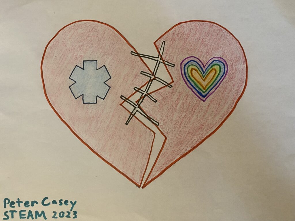
For my STEAM project I chose to research heart disease with a focus on how modern medicine utilizes care strategies to mitigate it’s effects. I chose to go a bit abstract with my art, in which a classic depiction of a heart shows how heart disease effects people from all walks of life but through constantly evolving practices, modern medicine seeks to repair the damage done.
There is a very good reason that throughout human history we have placed great importance on the heart. In everything from literature and science, to our art and music, the heart has become a symbol of life itself. And for good reason! The human heart truly is an amazing organ that our very survival depends on day in, and day out. It doesn’t get to take a break, there’s no going to sleep for a solid 8 hours of wonderful rest for the old ticker. No, the heart must continue it’s tireless work of maintaining life, and it’s to that tireless beat that the tune of our lives are played out, whether we’re aware of it or not.
However, like any organ or body system, the heart is subject to disease and/or failure. In fact, according to the (1) CDC, heart disease is the leading cause of death within the United States with 1 in every 5 deaths a result of heart disease in 2020. The vital and immediate role the human heart plays in life minute by minute means that when disease or failure of the heart occurs, life can end within an instant. While disease and failure of other body systems can produce similar results, none are quite so profoundly immediate as that of the heart. It is for this reason that science and modern medicine has gone to great lengths to produce definitive care strategies to maintain and restore the function of the heart. To better understand these strategies of care, we must first take a closer look at the heart and how it functions.
(2)The human heart is a highly evolved pumping structure that is actually 2 pumps in one, or a two-chambered pump. This is to ensure that pressure is maintained to both pulmonary and systemic circulation, a critical function of maintaining adequate oxygenation and perfusion of tissues. The layers of the heart starting from the outermost and moving in are the pericardium, the myocardium, and the endocardium. The coronary arteries and veins, which ensure perfusion of the heart itself, run through the epicardium along the surface of the heart with arterioles that run deep into the myocardium. It is these coronary arteries that can be highly susceptible to arteriosclerotic disease, becoming blocked and causing ischemia and/or death of the myocardium. The myocardium is the strong functional layer of myocardial or cardiac muscle cells. Cardiac muscle cells are specialized and unique cells that, among other things, have the ability to self excite which is also known as automaticity. This unique ability of automaticity means that the heart does not require neural control to continue pumping, and though neural and hormonal control is involved, the heart can maintain heart rate and perfusion without any outside control. This ability is a truly unique adaptation of the heart. Cardiac muscle cells have a number of other unique adaptations, like intercalated disks to ensure action potentials will be propagated throughout the heart, and a great many mitochondria (25-35% volume of the cell to be exact) that ensure the cardiac cells have the energy needed to maintain that tireless contraction. These great many mitochondria, means cardiac muscle cells have a greater dependence on Oxygen and are solely reliant on aerobic metabolism. Therefore even a small disruption of perfusion to the myocardium results in cell damage. The cells of the myocardium are further divided into two groups: 1. Contractile cells which, as you might expect, are responsible for the contraction of the heart, and 2. Pacemaker cells, namely the SA and AV nodes, which are non-contractile cells that spontaneously depolarize to create a depolarizing event that travels through the heart stimulating what we know as a heartbeat. The innermost layer of the heart is the endocardium, the smooth inner layer of the heart which lines the atria, ventricles and heart valves, and to which the chordae tendinea attach.
The heart is probably best known for it’s heartbeat, that “tha-thump” or “lub-dub” sound we all associate with a beating heart. The heartbeat represents what is known as the cardiac cycle, and to better understand it we’ll follow the heart through 1 complete cardiac cycle.
The cardiac cycle lasts about 0.8seconds and includes diastole, which is the filling phase, and systole, which is the contraction or ejection phase. Within systole we have the atrial systole, which lasts 0.1 seconds, and the ventricular systole, which lasts 0.3 seconds.
The cardiac cycle begins by the spontaneous depolarization of the SA node that is noted on an ECG (electrocardiograph) as the P wave. This precedes atrial contraction, which allows for the complete filling of the ventricles. The ventricles fill to 80% volume during diastole and the atrial contraction completes the last 20% of fill prior to ventricular contraction. It is a brief pause (0.1seconds) of the action potential at the AV node that allows for this atrial kick and complete filling of the ventricles. Once atrial contraction is complete, the atrioventricular valves (Tricuspid, and Mitral) close. At this point in the cardiac cycle all valves, both the atrioventricular and semilunar, are closed to allow for the isovolumetric contraction of the ventricles. The AV node will then pick up the action potential and next in the cardiac cycle is the Q wave of the QRS complex, which signals the beginning of ventricular systole and atrial diastole. During the QRS complex the atria will repolarize, as such the electrical signal of its depolarization is buried within the much larger electrical event of the QRS complex. Once the action potential of the QRS complex has propagated throughout the ventricles, the isovolumetric contraction of the ventricles will begin from the apex of the heart upward. Pressure within the ventricles will then increase with contraction until it exceeds pressure within the aorta and pulmonary arteries, leading to opening of the semilunar valves and onset of the ventricular ejection phase. This ejection phase occurs during the ST segment seen on an ECG and will show a sharp rise in pressure within both the ventricles and arteries, which can be felt as a pulse. The final event of the cardiac cycle is seen as the T wave on an ECG that signals repolarization of the ventricles and precedes isovolumetric relaxation of the heart. During isovolumetric relaxation all valves are closed and blood once again fills the atria, yet when pressure within the atria rises again from filling, the atrioventricular valves will open and passive filling of the ventricles will once again occur. This is the diastolic phase of the cardiac cycle, which sets the stage for the next cardiac cycle.
A more thorough understanding of the heart leads us next to the exploration of how we provide care for the heart when the delicate balance of electrical impulse, mechanical contraction and perfusion goes awry.
(3)The American Heart Association (AHA) is a leading authority on research and implementation of constantly evolving patient care strategies related to the cardiovascular system. Not least among the AHA’s tasks is training clinicians and the general public to recognize the signs of heart attack and to act quickly in an environment where time is everything, as the saying within the medical field goes “time is muscle”. This saying relates to the fact that myocardial cells are unable to convert to anaerobic metabolism in the absence of Oxygen, therefore are solely reliant on a constant supply of Oxygen from coronary circulation. If an interruption of perfusion to the myocardium occurs, ischemia and cell death is soon to follow. With the “time is muscle” concept in mind, the AHA has established a comprehensive strategy for the care of victims of heart disease in cardiac arrest that is called the “chain of survival”.
(4)The Chain of Survival focuses on 6 key elements or links, which can improve the chances of survival and recovery for victims of cardiac arrest. The 1st link in the chain is early recognition and activation of the emergency response system, this link in the chain emphasizes informational campaigns that teach the public about the signs of heart attack and cardiac arrest, followed by what actions to take should they witness such an event. The 2nd and 3rd links in the chain focus on high quality CPR and early defibrillation, with an emphasis on bystander CPR and placement of easy to use defibrillators in high traffic areas such as schools and shopping centers. (5) In fact studies have shown that when implemented correctly, bystander CPR and early defibrillation can have a profound impact on survival rates for out of hospital cardiac arrest. The 4th link in the chain involves rapid treatment using advanced resuscitation techniques by pre-hospital and in-hospital teams. Within this link time remains a key element. Both pre-hospital and in-hospital teams are trained to quickly deploy interventions like high performance CPR, rapid recognition of heart disease, followed by definitive treatment and restoration of heart function with fibrinolytics or coronary artery reperfusion therapy by a cardiac cath lab. The 5th and 6th links include post cardiac arrest care in a hospital setting, followed by recovery with additional treatment, rehabilitation, and ongoing observation. (6) Since implementation of the Chain of Survival in 2010, great efforts have been made across systems to strengthen each link within the chain, thereby improving the chances of survival and recovery of victims of heart disease and cardiac arrest.
In conclusion, it is easy to see why the human heart is deserving of it’s place of honor, and why doing everything we can to protect this strong yet delicate organ is a noble pursuit.
Peter Casey
STEAM 2023
References:
(1) Heart Disease Facts. (2022, October 14). Centers for Disease Control and Prevention. Retrieved March 25, 2023, from https://www.cdc.gov/heartdisease/facts.htm
(2) OpenStax Anatomy and Physiology Text book ISBN-13
(3) Emergency Cardiovascular Care. (2023). American Heart Association. Retrieved March 26, 2023, from https://cpr.heart.org/en/
(4) European Resuscitation Council. (2000). Part 12: From science to survival. Strengthening the chain of survival in every community. Resuscitation, 46, 417-30.
(5) Ritter, G., Wolfe, R. A., Goldstein, S., Landis, J. R., Vasu, C. M., Acheson, A., … & Medendrop, S. V. (1985). The effect of bystander CPR on survival of out-of-hospital cardiac arrest victims. American heart journal, 110(5), 932-937.
(6) Lin, H. Y., Chien, Y. C., Lee, B. C., Wu, Y. L., Liu, Y. P., Wang, T. L., … & Team, A. (2022). Outcomes of out-of-hospital cardiac arrests after a decade of system-wide initiatives optimising community chain of survival in Taipei city. Resuscitation, 172, 149-158.
Peter Casey’s 2023 steam project was great! It’s about the heart and how modern medicine and technology has advanced to increase care strategies. The leading cause of death in the United States is heart disease and 1 in every 5 deaths is from heart disease. When the heart fails, it’s immediate, unlike other organs where it would take more time to pass from organ failure. From the outermost to inside of the heart is the pericardium, myocardium, and the endocardium. Coronary arteries run through the epicardium and along the surface of the heart which in turn run deep into the myocardium. Those coronary arteries are very susceptible to arteriosclerotic disease where the arteries become blocked and cause death of the myocardium. The myocardium is home for these cardiac cells which allow the heart to continue pumping without any help from an outside source. This is just a little bit about the heart but this understanding of the heart and how it works allows heart disease scientists, like the American Heart Association, to be able to notice the signs of a heart attack sooner in order to reverse or fix what’s happening. They basically have a saying that “time is muscle” and work fast to find strategies to help those suffering from a heart attack. Peter also talks about the 6 chains of survival which are 1: early recognition 2 and 3: CPR and defibrillator control 4: advanced resuscitation 5 and 6: post cardiac care in the hospital or an intensive care unit. Overall, I learned a lot from Peter’s STEAM project and really enjoyed his project and artwork!