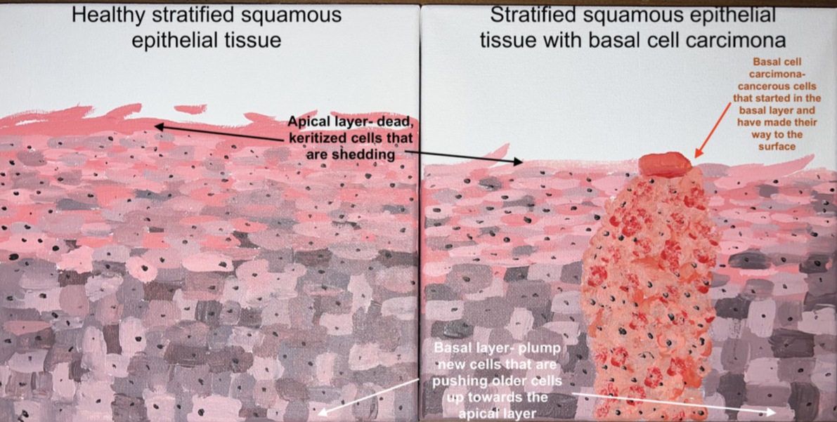
The objective I covered in this STEAM project is “Describe in depth each type of epithelial and connective tissue. Be able to describe their function and location.” I will specifically be discussing keratinized stratified squamous epithelium, its function, location, and issues that arise when the tissue is diseased. My paintings represent an example of healthy keratinized stratified squamous epithelial tissue and diseased keratinized stratified squamous epithelial tissue, respectively.
Keratinized Stratified squamous epithelium is a specific type of epithelial tissue. All epithelial tissues have polarity meaning they have a “top” and a “bottom”. The “top” is known as the apical surface which is the side that is exposed to a surface or cavity. The “bottom” is known as the basal surface, and it is the side that attaches to the basal lamina or the sheet that attaches this bottom to underlying cells. Epithelial tissues also all have specialized contacts that bind epithelial cells together, so they can fit closely. All epithelial tissues are supported by connective tissues and are avascular, but they do have nerve fibers. The connective tissue below the epithelium is what supplies nourishment to the epithelium since it is not able to do this on its own. Finally, all epithelial tissues are regenerative meaning they have the capacity to replace themselves. Epithelial tissue is found in many places that are physically abrasive; the skin, mouth, esophagus, etc. and this last characteristic is what keeps them going. The regeneration in the epithelial cells is stimulated when lateral specialized contacts are broken and when there is a loss of apical-basal polarity.
Epithelial tissue is classified by the shape of its cells and the number of cell layers. Stratified epithelium has two or more layers of cells. Squamous cells are flattened, and scale like in shape. So, stratifies squamous epithelium means that the tissue would have multiple layers of flattened cells. It’s also important to note that the shape is specifically what is present in the apical layer of cells. You will notice in the paintings that both seem to have cells that are plumper than you would think when discussing squamous cells, some may even seem cuboidal or columnar and that’s normal. If you look these cells are towards to basal surface and as you move up to the apical surface the cells will be flattened and like the classic squamous cell you would think of. The term keratinized is referring to the specific “dry” stratified squamous epithelial tissue found in the skin. The cells that seem plumper towards the basal layer are called
Seaira Jacques Anatomy & Physiology I STEAM Project Essay
basal cells and are active in producing more cells. The apical cells also known as surface cells are dead and flat (notice the flakes at the apical surface of the paintings). Stratified squamous epithelial tissue make up the body’s first layer of defense against foreign objects outside of the body. The multiple layers that make up the stratified squamous epithelium protect underlying tissues from harm. The surface cells that are dead can flake off without causing issues to the underlying tissue while the basal cells help to reproduce new cells to make up for the lost cells.
Basal Cell Carcinoma is a cancer that specifically affects Keratinized Stratified Squamous Epithelial Tissue. This cancer develops as a result to exposure to UV rays (most commonly chronic sun exposure. The UV exposure damages the DNA within the basal cells. (Badri, McDaniel, Steel, 2022) When the basal cells that are responsible for organizing reproduction of the epithelial cells are damaged this can cause a build of cells. This buildup of epithelial cells is what presents itself as what the public refers to as skin cancer; spots, bumps, scaley patches or discoloration. (Oh, Stark, Reichrath, 2020) There are different forms of skin cancer that present differently, and even different cases of basal cell carcinoma can present differently in each patient. The common ground that is shared with all basal cell carcinomas is that it is directly affecting the function of the keratinized stratified squamous epithelium. The diseased DNA causes the basal cells to soften and overproduce cells creating tumors. The diseased tissue is then not able to serve its purpose of protecting underlying tissue if the tumor grows large enough. This is because the diseased cells don’t keep the sheet of stratified squamous epithelium together. Basal Cell Carcinoma is usually slow growing and is not usually deadly due to how slow it grows. It is also in a location (keratinized stratified squamous) where once the tumor and diseased tissue is removed the healthy epithelial tissue can regenerate and reform the specialized connections. There may be evidence of the basal cell carcinoma in the form of a scar, but that tissue is not diseased and is able to perform its functions properly without issue. (Fiore, Krajnc, Kevorse, Pasolli, Shcarttsman, Fuchs, 2020)
Seaira Jacques Anatomy & Physiology I STEAM Project Essay
Citations:
Fiore, V. F., Krajnc, M., Quiroz, F. G., Levorse, J., Pasolli, H. A., Shvartsman, S. Y., & Fuchs, E. (2020). Mechanics of a multilayer epithelium instruct tumour architecture and
function. Nature, 585(7825), 433–439. (Peer-reviewed)
McDaniel B, Badri T, Steele RB. (2022) Basal Cell Carcinoma. Treasure StatPearls 1-13
Oh, S. T., Stark, A., & Reichrath, J. (2020). The p53 Signalling Pathway in Cutaneous Basal Cell Carcinoma: An Immunohistochemical Description. Acta dermato-venereologica, 100-106 (Peer- reviewed)
Seaira talks about Keratinized stratified squamous epithelium, its function, location, and issues that arise when the tissue is diseased. The objective covered is “ Describe in depth each type of epithelial and connective tissue. Epithelial tissue is classified by the shape of its cells and the number of the cell layer. Keratinized Stratified Squamous epithelium is a specific type of epithelial tissue. All epithelium tissues have polarity meaning they have a “top” and a “bottom”. All epithelial tissues are regenerative meaning they have the capacity to replace themselves. The regeneration in the epithelial cells is stimulated when lateral specialized contacts are broken and when there is a loss of apical basal polarity.
Seaira also talks about a type of cancer that affects keratinized Stratified Squamous Epithelial Tissue called “ Basal Cell Carcinoma”. This cancer develops as a result to exposure to UV rays. The UV exposure damages the DNA within the basal cells. When the basal cells that are responsible for organizing reproduction of the epithelial cells are damaged this can cause a build of cells. Basal Cell Carcinoma is usually growing slowly and is not usually deadly due to how slow it grows.It is also in a location where once the tumor and diseased tissue is removed the healthy epithelial tissue can regenerate and reform the specialized connections.
Seaira’s painting represents an example of healthy keratinized stratified squamous epithelial tissue and diseased keratinized stratified squamous epithelium tissue.
It’s a great work of art! You can definitely tell Seaira spent lots of time doing research and painting. I love it!