For my project I wanted to explore the topic of epidurals. The objectives that I find relevant to this topic include, compare, and contrast the central nervous system, Identify the 7 functions of the bone, know the parts of the bone and their shape. When a woman gets an epidural, she gets it to alleviate the pain of contractions and such from childbirth. For an epidural, there is a shot given to numb the area and then another shot goes into a womans back to relieve the pain in her lower body, let’s dive deeper into that.
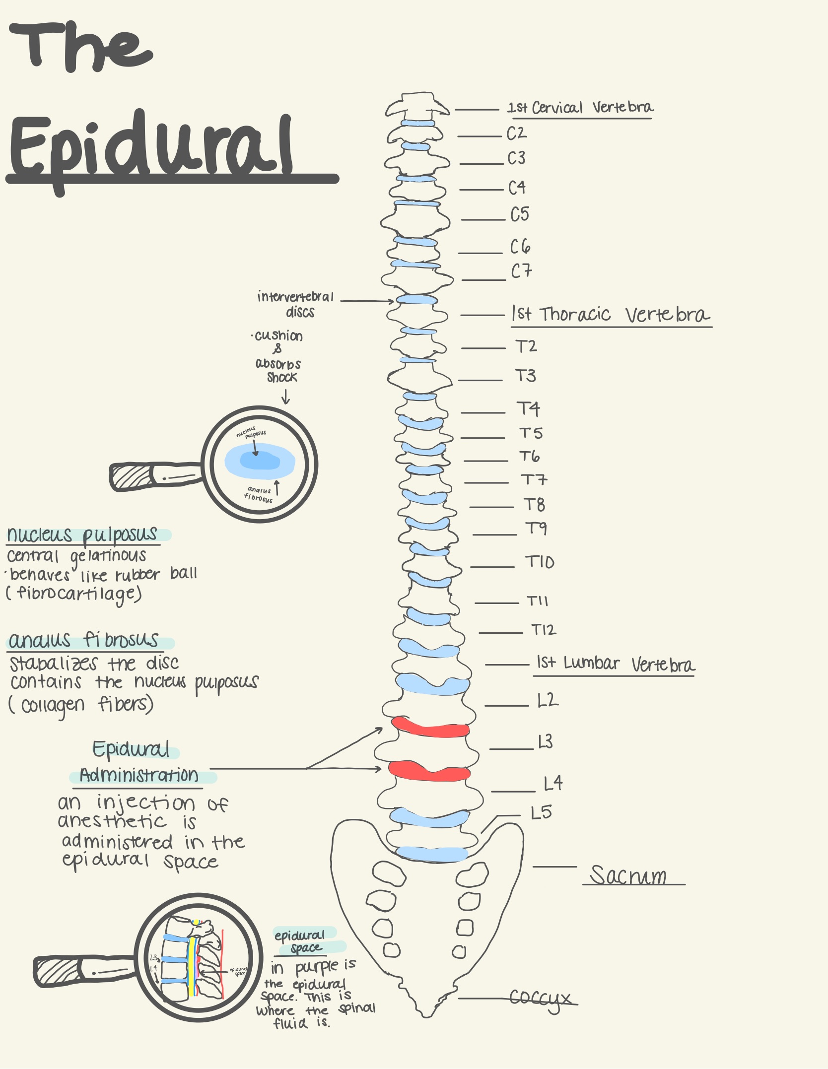
For my creative component I drew a vertebra which consist of 24 vertebrae bones that extends from the skull to the pelvis and is the upper bodies main support. The three sections of the vertebrae are the cervical vertebra, thoracic vertebra, and the lumbar vertebra. These sections that make up the vertebra play a huge role in protecting the spinal cord. One of the functions of bone is protection, and the vertebra protects the spinal cord. The vertebrae bones are considered irregular bone because of their odd shape and in between each vertebrae bone is intervertebral discs. These discs make it so there is cushion in between these bones and that they don’t grind against each other. Another key thing is that because of their structure they can absorb shock, the intervertebral discs consist of two parts, the nucleus pulposus which is the “nucleus” or the inside of the discs which is gelatinous and is like a rubber ball. Then there is the annulus fibrous which is what stabilizes the discs and is what contains the nucleus pulposus which is collagen fibers which lets us know that it’s very sturdy. Epidurals occur between the L3 and the L4 bones on the vertebra. When we take a closer look into the vertebrae itself there’s the spinal cord but there is also the epidural space.
Getting even deeper into the components of an epidural, looking into the spinal cord it’s a part of the central nervous system along with the brain. There are many parts of the spinal cord anatomy but one of the most important parts is the epidural space. In my diagram I drew a close-up of where the epidural space would be. When a healthcare provider inserts the needle into your back it goes through skin and then into your epidural space where there is consistent anesthetic going into your spinal fluid to relieve pain. To reduce the amount of pain the epidural medicine that goes into your spinal fluid blocks nerves which affects your lower body. The epidural relieves some pain, but it doesn’t relive it all. This is because it’s important that the woman still can function and push to get her baby out.
The digital art that I wanted to make a poster that you would see in maybe a hospital, so it’s informational. I made is a structure of the vertebra with the components and it also includes the coccyx and the sacrum. I did a close-up of the interverbal discs to show which section is the annulus fibrous and the nucleus pulpous. I also did a close-up of the spinal cord and where the needle is inserted in the epidural space.
References:
Berillis, P., Panagiotopoulos, N., Boursiaki, V., Karapanagiotidis, I. T., & Mente, E. (2015, May 8). Vertebrae length and ultra-structure measurements of collagen fibrils and mineral content in the vertebrae of Lordotic Gilthead seabreams (sparus aurata). Micron. Retrieved November 10, 2022, from https://www.sciencedirect.com/science/article/pii/S096843281500075X
Editor. (2021, December 9). What is an epidural? American Pregnancy Association. Retrieved November 10, 2022, from https://americanpregnancy.org/healthy-pregnancy/labor-and-birth/what-is-an-epidural/
Epidural space. Epidural Space – an overview | ScienceDirect Topics. (n.d.). Retrieved November 10, 2022, from https://www.sciencedirect.com/topics/medicine-and-dentistry/epidural-space
This project is done exceptionally well. Right off the bat, the diagram is neat, concise, and very accurate, it’s very easy to understand and it includes some essential vocabulary and definitions of the different parts of the anatomy of the spine. The labeling of the other parts of the spine is very neat and perfectly displays all the different individual discs/bones and clearly defines the different sections of the spine all the way down to where exactly the epidermal is administered. labels on the side of the smaller parts and the descriptions that give context are also done very well, they’re very simple and informative.
Within the description of the abstract, the objective is clearly stated and related back to the project’s purpose. The purpose of this project is to show where epidural injections are administered and how exactly it works. They begin with giving a overal summary of the spine starting with how it had 24 vertebrae and is the “the bodies main support”, following up with defining the different sections of the spine (cervical, thoracic, and lumbar vertebrae). Additionally, they describe the discs within the spine and how they provide cushioning between the vertebrae. Lastly the describe the components of the discs and then state where in the so9e the epidural is administered.
They also go on to describe how it is administered. By referencing the abstract, they can accurately describe where a healthcare worker can insert the needle and where exactly the needle will end up. It also helps them explain how the epidural releases anesthetic into the spinal fluid which will then relieve the pain while allowing the woman to still push because only the nerves in the lower body are affected. Overall this project is excellent and I learned something that I otherwise may not have gone out of my way to learn about.