
The topic of my STEAM Project is accessory navicular syndrome. The two objectives I’ve represented through my project are 1) know the stages of bone development and repair and 2) know the parts of the bone and their shape. Accessory navicular syndrome is when a person is born with an extra bone in their foot just outside of their navicular bone. Both the navicular and accessory navicular bones are irregular shaped bones, with the accessory bone being a small irregular bone. These bones have a unique curved shape and don’t have any real sort of structure to them.
A lot of people won’t even realize they have this extra bone, because if it isn’t too disturbed in a person’s youth the accessory bone will fuse to the navicular bone. This bone fusion happens at the ages of 9-11 years old and after this fusion happens even if a person had this extra bone, they won’t feel any additional pain if they roll their ankle. Their feet would be completely normal and developed, but perhaps have a larger medial region on their foot. This bone fusion and future development won’t happen if the foot is distressed prior to the ages of 9-11. Therefore, accessory navicular syndrome is most common in young athletes who are constantly putting harsh pressure on their feet. I was personally born with accessory bones in both of my feet, but my right accessory bone caused me the most irritation to the point where I had to have it surgically removed. When I was in middle school, everytime I would even slightly twist my ankle I wouldn’t be able to walk for a few days. My extra bone would move with my foot as it twisted, which caused me so much pain. After I was healed I continued to play sports and in five or six months I would twist my ankle and the extra bone would move again. When I got to high school the possibility of bone fusion was out of the question. My accessory bone had moved too much and I was planning on playing basketball in college, so the only option was surgery.
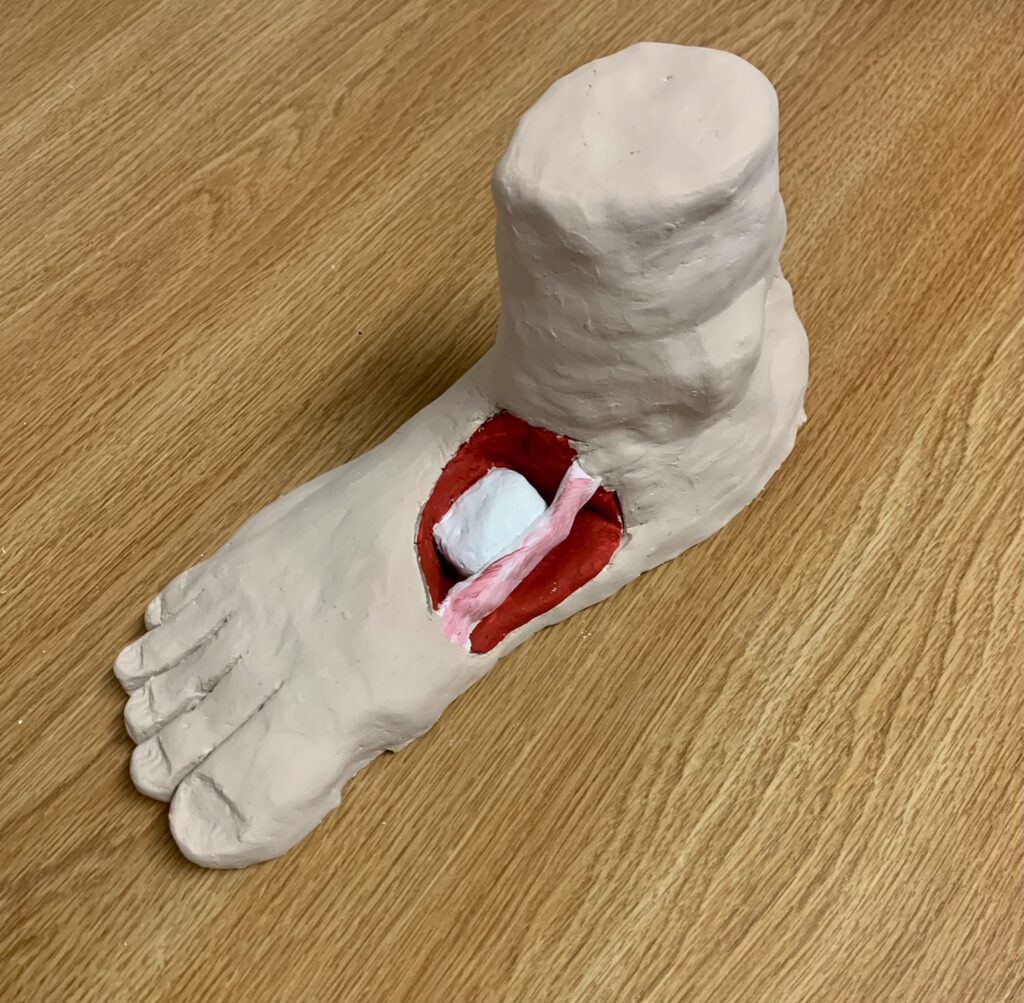
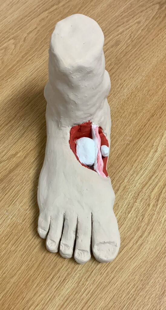
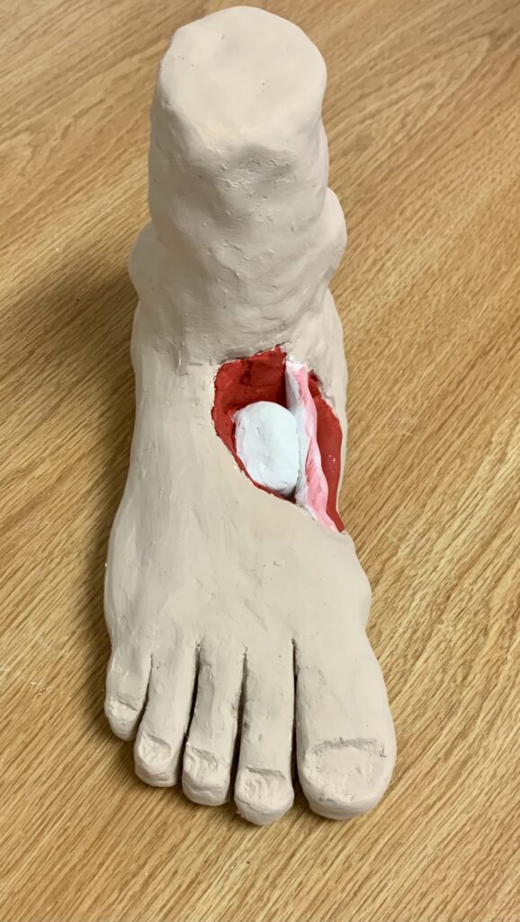
I made a clay model of a foot to resemble what my foot looked like pre surgery (with accessory bone) and post surgery (accessory bone removed). As you can see on the model, there’s a larger navicular bone on the interior of the foot, then a tendon attached to that bone, followed by the accessory bone attached to that tendon. This tendon is called the posterior tibial tendon. This tendon is a big reason I felt so much pain whenever I twisted my ankle. As my ankle rolled, the tendon would bend with my ankle, and my extra bone would move with it. In fact, my surgeon told me he would have to detach the accessory bone from my tendon, which would add time to my recovery period. However, during the surgery my accessory bone was positioned in a way where he didn’t have to touch the tendon and he simply removed the extra bone. My clay foot model also presents the foot to be extremely flat, which is also a sign of accessory navicular syndrome. One of the functions of the posterior tibial tendon is to hold up the arch of your foot, so this arch may drop if an extra bone is disrupting the tendon from doing its job. After this surgery I was instructed by my surgeon to hardly ever walk barefoot. My tendon will never be the same after surgery, so I constantly have to walk with arch support slippers and put arch inserts into all the shoes I wear, so as to not have my arch drop even lower due to my loose posterior tibial tendon.
References
- Jegal H. (2016). Sage Journals. Accessory Navicular Syndrome in Athlete vs General Population. 37(8). https://doi.org/10.1177/1071100716644791
- Strayhorn G. (1982). The Journal of Family Practice. The Symptomatic Accessory Navicular Bone. 15(1). 59-64. https://cdn.mdedge.com/files/s3fs-public/jfp-archived-issues/1982-volume_14-15/JFP_1982-07_v15_i1_the-symptomatic-accessory-navicular-bone.pdf
- Parekh S.G. (2019). OrthoInfo. The accessory navicular: “I have an extra bone in my foot?”. https://orthoinfo.aaos.org/en/diseases–conditions/ortho-pinion-accessory-navicular/