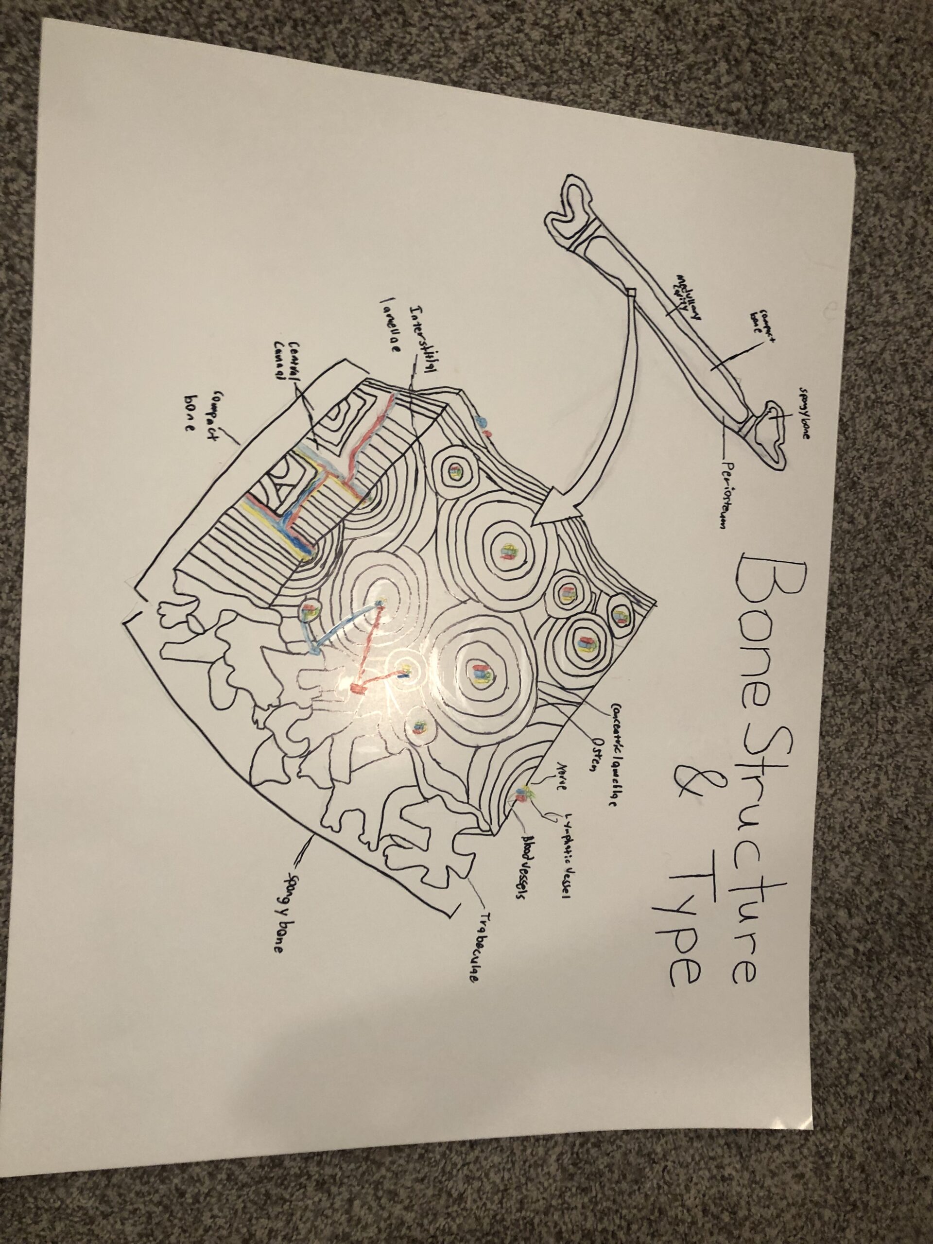
The following is a scientific concept presentation expressed through art along with this STEAM project statement. The project chosen is the human compact bone, also called cortical bone, and reviews its structure and use. The drawing with this written statement displays a femur bone and an expanded diagram of the inside bone structure. The expanded structure shows eleven intercellular materials. According to Kozielski et al (2011), bone tissues make up the main component of the skeletal system. Bone has a unique type of connective tissue created from certain types of cells such as osteognic cells, osteoblasts, and osteocytes. The mineral component of bone includes apatite crystals, calcium salts, and water. Bone structure strength depends on these mineral components. Organic components are made up from collagen fibers. These determine elasticity and toughness of the tissue. Schlesinger et al (2020) note that bone and other connective tissues are isolated by a layer of osteoblasts. This helps in the formation of compact and strong bone structure.
The bone drawing on the left side of the poster displays the following from outside to inside structure: periosteum, medullary cavity, compact bone, and spongy bone. The expanded drawing displays the internal structure. Inside the compact bone is the Periosteum (outer fibrous layer and inner osteogenic layer), interstitial lamellae, perforating canal, central canal, blood vessels, lymphatic vessel, nerve, trabeculae, osteon, and concentric lamellae. The role of the periosteum is to connect tissue that covers the outer surface of the bone. Interstitial lamellae is between the osteons which are a central canal surrounded by concentric rings called lamellae. These vessels interconnect through the perforating canals with vessels on the surface of the bone. Perforating canals allow blood vessels and lymphatic vessels to link with nerves in the central canals. Trabeculae is the porous form of bone tissue organized into a network of rods and plates filled with bone marrow inside the medullary cavity (Lumenlearning, 2021). Bones are the strongest connective tissue which provides support, protection, and movement for the body. Bone properties must include collagen and mineral crystals to remain strong and yet flexible. Transverse tissue of long bones is arranged of osteocytes in concentric circles around a central canal. Since bone is a vascularized tissue, it can recover from injuries in a short period of time (OpenStax, 2021).
Compact bones store and release calcium to the body when needed. According to Wei and Sun (2018), a common compact bone disease known as osteoporosis is due to aging related changes in bone colles, extracellular matrix and molecules causing bone loss and fractures. The disease affect bone mass density. Skeletal components cells include osteoblasts, osteoclasts, osteocytes, and bone marrow cells play a role in skeletal development, maintenance, and the pathogenesis (development of a disease) osteoporosis. Senescence of cells lead to osteoporosis in aging people. However, Sunyecz (2008) reposts that the use of calcium and vitamin D are part of a sound maintenance of osteoporosis. The author notes that about 1.5M osteoporotic fractures occure in women and in in the U.S. The U.S. Surgeon General reports one of two women over the age of 50 will have an osteoporosis-related fracture in their lifetime. Calcium and vitamin D studies indicated a daily recommended dietary supplement for strong bone maintenance.
Conclusion
This brief presentation provided an overview of the compact bone structure, the internal structure, and how they are interconnected. The compact bone is the strongest bone in the body which we rely on for strength, flexibility, and movement. However, when we age, we become susceptible to osteoporosis. A daily supplementary intake of calcium and vitamin D are needed for healthy bone maintenance. If we do experience a bone breakage, the compact bone is a vascularized tissue, and has the ability to heal quickly.
References
Kozielski, M., Buchwald, T., Szybowicz, M., Błaszczak, Z., Piotrowski, A., & Ciesielczyk, B. (2011). Determination of composition and structure of spongy bone tissue in human head of femur by Raman spectral mapping. Journal of materials science. Materials in medicine, 22(7), 1653–1661. https://doi.org/10.1007/s10856-011-4353-0
LumenLearning. (2021). Anatomy and physiology I: Module 7 – Bone tissue and the skeletal system. https://courses.lumenlearning.com/hccs-ap1/chapter/bone-structure/
OpenStax. (2021). Anatomy and physiology. https://openstax.org/books/anatomy-and-physiology/pages/4-3-connective-tissue-supports-and-protects?query=compact%20bone&target=%7B%22index%22%3A3%2C%22type%22%3A%22search%22%7D#fs-id1517273
Schlesinger, P. H., Blair, H. C., Beer Stolz, D., Riazanski, V., Ray, E. C., Tourkova, I. L., & Nelson, D. J. (2020). Cellular and extracellular matrix of bone, with principles of synthesis and dependency of mineral deposition on cell membrane transport. American Journal of Physiology: Cell Physiology, 318(1), C111-C124. https://doi.org/10.1152/ajpcell.00120.2019
Sunyecz J. A. (2008). The use of calcium and vitamin D in the management of osteoporosis. Therapeutics and clinical risk management, 4(4), 827–836. https://doi.org/10.2147/tcrm.s3552
Wei, Y., & Sun, Y. (2018). Aging of the bone. (pp. 189-197). Springer Singapore. https://doi.org/10.1007/978-981-13-1117-8_12
Bones make up the main component of the skeletal system, which we rely on for strength, flexibility, and movement. They are each made up of connective tissue created from osteognic cells, osteoblasts, and osteocytes. The mineral components of bone are apatite crystals, calcium salts, and water (mineral components determine strength of structure). Organic components are made up from collagen fibers which determine the elasticity and strength of the tissue. Bones are lined with a layer of osteoblasts that help in the formation of new compact bone.
The layers shown in the drawing to the left from superficial to deep are periosteum, medullary cavity, compact bone, and spongy bone. The drawing to the right is expanded, showing inside the compact bone is the Periosteum (outer fibrous layer and inner osteogenic layer), interstitial lamellae, perforating canal, central canal, blood vessels, lymphatic vessel, nerve, trabeculae, osteon, and concentric lamellae. Compact bones store and release calcium to the body when needed. A common bone disease called osteoporosis ( most common in women over 50 yrs), is due to age-related changes in bone colles, extracellular matrix and molecules causing bone loss and fractures; osteoporosis affects bone density. Luckily, the use of calcium and vitamin D are part of a sound maintenance of osteoporosis.