The Contraction “Cycle”
Kyle McClendon
University of Alaska Fairbanks
Abstract
A short description of the Image of the Contraction Cycle presented and worked on by Kyle McClendon, in ownership of the University of Alaska Fairbanks.
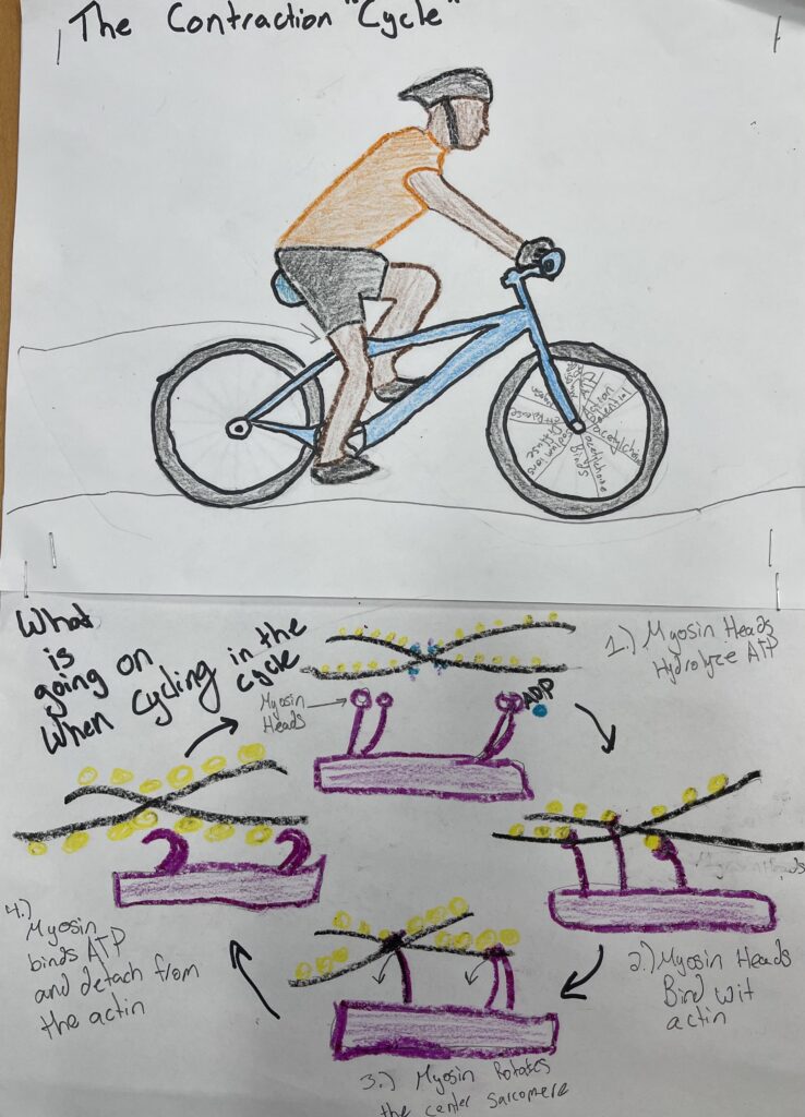
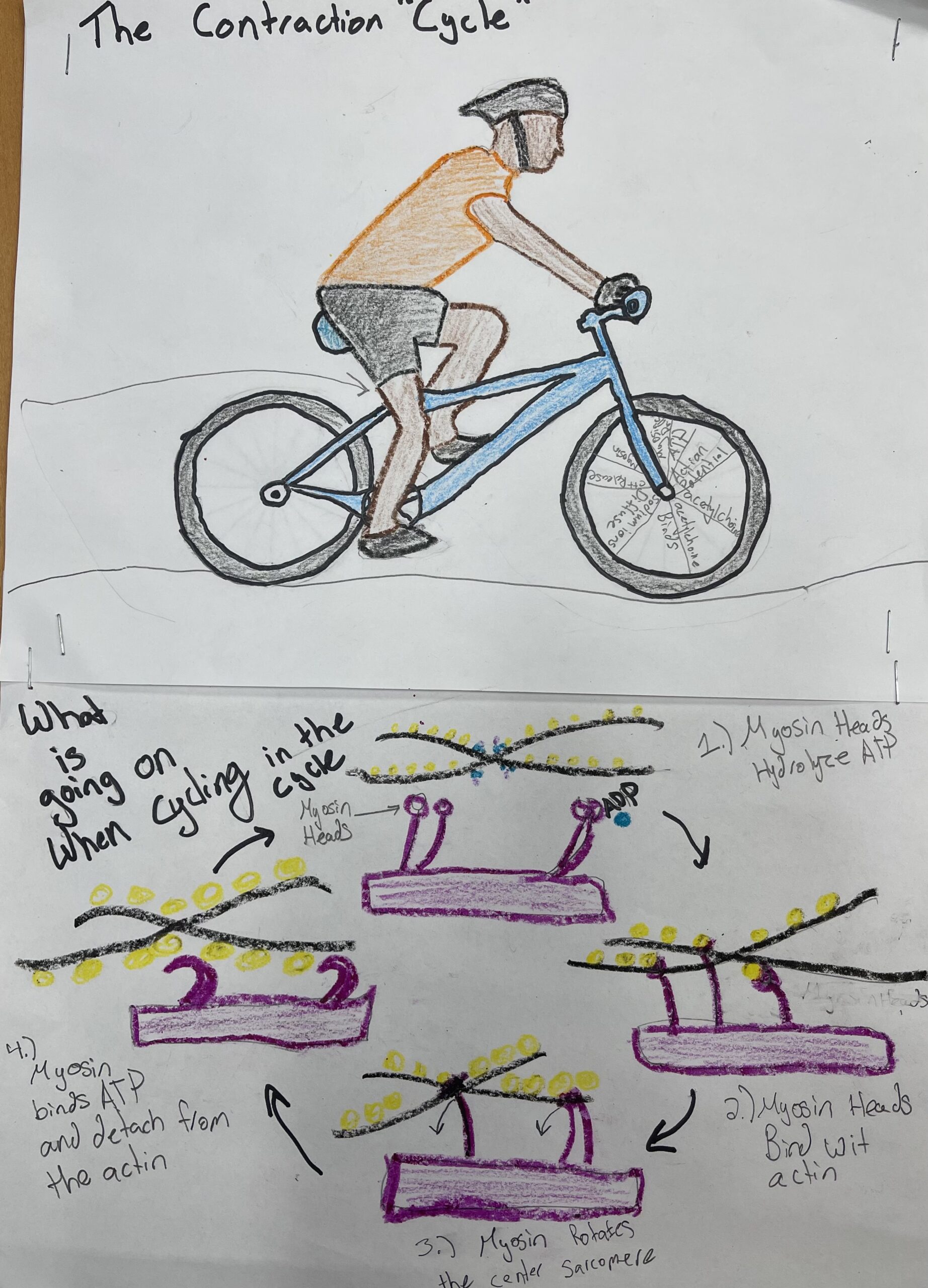
The Contraction “Cycle”
The drawing of the contraction “Cycle” is to represent and illustrate the contraction cycle of myosin heads combining between the Actin strands. This illustration shows an athletic and fits person riding a bike with their legs doing most of the power to produce energy to move the bike.
The Cycle
As illustrated in the drawing, the biker is propelling with their feet, legs, and core muscles and moves on flat ground. The biker is utilizing most of the muscles in the body in order to produce energy to propel the bike. The biker’s muscles are also contracting and releasing in order to provide contraction to each muscle group that is needed to propel the bike. On the front spokes of the wheel, there are eight spokes to illustrate the eight steps of muscle contraction and release.
The steps1
the four main steps that are required to provide muscle contraction to the human body include two main parts. These parts are the myosin heads and the Actin strands that utilize each other in order to produce a muscle contraction. The main ingredient in order to produce a muscle contraction is the myosin heads and the ADP with calcium. As calcium is released, the myosin heads are able to grasp the actin strands and create a muscle contraction. These four steps are utilized in all muscle contractions. R step one is the hydraulic sized ATP becomes oriented and energized via the myosin heads. Step two requires the myosin heads to fasten or bind to the actin. This creates a cross-bridge between the myosin and actin. Step three requires the myosin to cross-bridge and latch onto the actin and move toward the sarcomere. This is also considered a power stroke. The fourth step is the myosin heads fastened to the ATP, forming the cross-bridge to detach from the actin. The final step is for the calcium to be pumped out of the muscle in order to have released. If there is not calcium out of the muscle, it’ll cause rigor mortis, which is commonly seen in fatal accidents. People that are not alive will have rigor mortis as the body is not able to release the calcium out of the muscles and not able to release the muscle. In this case, ATP is doing the mechanical work of combining muscle and energy.
The reason for choosing this project. The reason that I chose to use a cyclist as an example for the contraction cycle is to show that the muscle contraction of a cyclist can correlate to that of the contraction cycle. The contraction “Cycle” was a good play on words in order to have a better understanding of how this cycle is completed throughout the body. Kyle McClendon likes to avid downhill bikes and utilizes each muscle group as needed in order to propel the bike down the hill or up the hill. This is how it was relatable to Kyle McClendon needed to have a better understanding of how the contraction cycle has functioned.
References
10.3 Muscle Fiber Contraction and Relaxation – Anatomy and Physiology. OpenStax. (n.d.). https://openstax.org/books/anatomy-and-physiology/pages/10-3-muscle-fiber-contraction-and-relaxation.
Boundless. (n.d.). Boundless Biology. Lumen. https://courses.lumenlearning.com/boundless-biology/chapter/muscle-contraction-and-locomotion/.
Their, Butler, & Lewis Hole’s Human Anatomy 10th edition McGraw Hill, Boston 2004. (2016). Muscular System: Stages of A Muscle Contraction. A. http://legacy.owensboro.kctcs.edu/gcaplan/anat/notes/api%20notes%20j%20%20muscle%20contraction.htm.