Bone regeneration post fracture. This includes natural growth as well as assisted growth with rods and screws.
One Comment
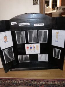
This information board by William shows X-Ray images of different bones during regeneration and growth. Each side of the board has drawn images of bones. There is a neonatal bone on the left. Neonatal bones are the developing bones of a newborn. In the picture, you can see a great amount of hyaline cartilage. Hyaline cartilage is flexible and resistant to damage. In the middle of the board is the regeneration process. The neonatal bone has intact epiphyseal cartilage that allows room for growth. When a bone initially breaks, a large hematoma forms. There is a lot of swelling and pain. Then an internal and external callus forms along with blood vessels. Then a bony callus of spongy bone forms. The bone gets stronger and stronger with increased blood and marrow to the site. Lastly, the medullary cavity is reformed, and outer regions of spongy bone turn to compact bone. On the right of the board are an X-Ray image and drawing of an adult bone. Also shown here is what the bone looks like with rods and screws. Rods and screws are used in more severe fractures, like compound fractures. These tools are useful in avoiding deformities, lengthening, and adding support. The rods would be surgically placed in the medullary cavity from the proximal or distal portion of the bone head. Rods are secured by screws. Rods can temporarily damage marrow stores. Ports can be inserted into the rods to provide drainage and diminish the chances of infection and sepsis.

This information board by William shows X-Ray images of different bones during regeneration and growth. Each side of the board has drawn images of bones. There is a neonatal bone on the left. Neonatal bones are the developing bones of a newborn. In the picture, you can see a great amount of hyaline cartilage. Hyaline cartilage is flexible and resistant to damage. In the middle of the board is the regeneration process. The neonatal bone has intact epiphyseal cartilage that allows room for growth. When a bone initially breaks, a large hematoma forms. There is a lot of swelling and pain. Then an internal and external callus forms along with blood vessels. Then a bony callus of spongy bone forms. The bone gets stronger and stronger with increased blood and marrow to the site. Lastly, the medullary cavity is reformed, and outer regions of spongy bone turn to compact bone. On the right of the board are an X-Ray image and drawing of an adult bone. Also shown here is what the bone looks like with rods and screws. Rods and screws are used in more severe fractures, like compound fractures. These tools are useful in avoiding deformities, lengthening, and adding support. The rods would be surgically placed in the medullary cavity from the proximal or distal portion of the bone head. Rods are secured by screws. Rods can temporarily damage marrow stores. Ports can be inserted into the rods to provide drainage and diminish the chances of infection and sepsis.