
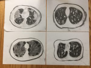
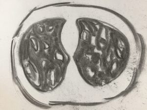
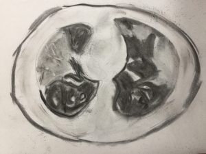
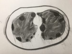
Top: COVID-19 lung damage (left), parasitic lung damage (right)
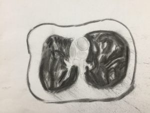
Bottom: COPD (left), healthy lung (right)
Top: COVID-19 lung damage (left), parasitic lung damage (right)
Bottom: COPD (left), healthy lung (right)
Kyndall Powers
There are several ways to diagnose lung issues, and a common way we keep hearing about lately due to COVID-19 is a computerized tomography scan (CT). I recreated CTs from healthy lungs, then lungs impacted by COVID-19, chronic obstructive pulmonary disease (COPD), and parasitic infection. I used homemade charcoal on a lightweight canvas to recreate these images.
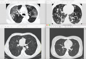
A transverse CT of your thorax uses low-dose x-ray to provide a clearer picture of your lungs over a standard chest x-ray and was first employed in the 1970s (Goldman 2007). The first thing to notice on the CT scans is that the left and right lung are not the same shapes. The left lung has a notch that accommodates the heart. Different tissue densities will show up as varying grey-scale. Air is black, normal lung parenchyma lighter black compared to air. and mucus is white. Due to this variation, contrast is not needed to view the lungs via CT.
In the COVID-19 lungs have what is called a ground-glass opacities (GGO) and crazy-paving pattern that appears as a light-ish grey in large patches of the lung and is indicative of pneumonia (Pan et al. 2020). Over time, the ground class and crazy-paving pattern are partially absorbed though the long-term effect of COVID-19’s lung damage is still mostly unknown (Pan et al. 2020).
Chronic obstructive pulmonary disease (COPD) is a leading cause of death in the world (Vestblo et al. 2013). It is characterized by the reduction of expiratory airflow that is a result of anatomical lesions (Vestbo et al. 2013). Chronic inflammation interrupts regular repair, which leads to fibrosis (Vestbo et al. 2013). The dark patches on the CT of lungs impacted by COPD is trapped air.
Parasitic lung damage can be caused by protozoa, nematodes, and trematodes (Kunst et al. 2011). The CT scan depicted in my project is the result of damage from schistosomiasis, a tropical trematode that infects roughly 200 million people worldwide (Kunst et al. 2011). As it passes through the lungs, it can cause pneumonitis, which is the inflammation of the alveoli (Kunst et al. 2011). In a CT, ground-glass opacification, which is also seen in COVID-19, appear along with nodular changes (Kunst et al. 2011).
Literature Cited
Goldamn, L. (2007). Principles of CT and CT Technology. Journal of Nuclear Medical Technology. 35(3), 115-128.
Kunst, H., Mack, D., Kon, O. M., Banerjee, A. K., Chiodini, P., & Grant, A. (2011). Parasitic infections of the lung: A guide for the respiratory physician. Thorax, 66(6), 528— 536.
Pan, F., Ye, T., Sun, P., Gui, S., Liang, B., Li, L., Zheng, D., Wang, J., Hesketh, R.L., Yang, L. and Zheng, C.. (2020). Time course of lung changes on chest CT during recovery from 2019 novel coronavirus (COVID-19) pneumonia. Radiology, 200370.
| Vestbo, J., Hurd, S.S., AgustÃ, A.G., Jones, P.W., Vogelmeier, C., Anzueto, A., Barnes, P.J., Fabbri, L.M., Martinez, F.J., Nishimura, M. and Stockley, R.A (2013). Global strategy for the diagnosis, management, and prevention of chronic obstructive pulmonary disease: GOLD executive summary. American journal of respiratory and critical care medicine, 187(4), 347-365. |
Computerized tomography or CT scans are a common diagnostic tool for lung issues and disease. CT scans use a low-dose x-ray to depict a clear image of the lungs. The air in the lungs appear black, normal lung parenchyma appears as a lighter black, tissues appear as a variety of greys depending on their density, and mucus appears white. A CT scan of COVID-19 affected lungs shows ground-glass opacities and crazy-paving patterns also seen in other lung diseases. CT scans of COPD affected lungs reveal dark patches of trapped air. Lastly, parasitic lung infections also show ground-glass opacities, similar to that in COVID-19, and followed by nodular changes.