 Posted by
Course Objective: Know the tissues/cells and functions of the following: stomach, pancreas, liver, gallbladder, large and small intestine, and esophagus, to these functions.
Colorectal cancer has been a topic that is more present in social media, with reminders to keep an eye on your health, stool, and stay up to date with colonoscopies. People are becoming more aware that colorectal cancer (CRC) is the third most diagnosed cancer, and the second most common cause of cancer-related deaths worldwide (Duan et al., 2022). CRC usually occurs in people aged 45 or older. It is found that family history, environmental factors, lifestyle, diet high in fat and protein, smoking, and alcohol consumption can increase the risk of colon cancer (Hossain et al., 2022 & Rice University, 2022, p. 1028)
The colon is found within the large intestine between the cecum and rectum (Larson, Unit 13 slide 86). The mucosa, the inner lining of the colon, is made up of simple columnar epithelial tissues. These tissues have microvilli and cilia that help with the absorption and secretion of mucus and enzymes (Larson, Unit 3 slide 19). Followed by submucosa that is made up of dense irregular connective tissue and helps resist tension from different directions (Larson, Unit 3 slide 48) In the early stages, the mucosa and submucosa are the first to be hit by CRC through “… hyperplasia, atypical hyperplasia (mild, moderate, severe) and adenomas, that can eventually develop into carcinoma” (Duan et al., 2022). This is the result of cancerous factors like chromosomal instability and genetic mutations, which are changes in DNA structure that lead cells to become cancerous (Duan et al., 2022).
Colorectal cancer spreads and metastasizes (the spread of cancer through tumors) through four pathways: local invasion, lymphatic metastasis, hematogenous metastasis, and implantation and metastasis. Local invasion is when the tumor infiltrates the precancerous location and adjacent structures. Lymphatic metastasis is when (pre)cancerous cells use the lymphatic system through the mucous membrane to reach lymph nodes and spread, causing lymph node metastasis. The hematogenous metastasis happens when the cancer cells spread through the blood vessels, targeting the liver and lungs. Lastly, implantation and metastasis are when the cancerous cells fall off the mucous membrane and implant in the abdomen and pelvic cavity (Duan et al., 2022). It takes up to eighteen years to develop a polyp and invasive cancer, with an average of nine years for metastasis to form (Hossain et al., 2022).
The right side of the colon mainly absorbs water and some nutrients, while the left side stores and excretes feces. All while secreting gastrointestinal hormones and alkaline mucus substances to help regulate gastrointestinal hormones and act as a protective barrier and lubricant (Duan et al., 2022). With colorectal cancer present, bowel movements change due to the tumor that has grown and disrupts normal function. Bleeding can occur due to tumor damage to the inner lining of the colon.
Colorectal cancer is slow in developing over the span of several years to decades (Hossain et al., 2022). This is why preventative care is crucial in catching CRC early on by seeing if there is abdominal discomfort, changes in stool, blood in stool, unexplained progressive weight loss, or abdominal mass (Duan et al., 2022). When these symptoms are noticed, doctors will do further examinations. When cancer is found, it can be treated with colonoscopies, chemotherapy, targeted therapy, or gene therapy, to name a few (Hossain et al., 2022).
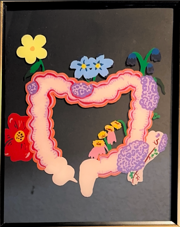
In my art piece, the colon is separated into five parts to show the progression of a healthy colon to the four stages of cancer from the cecum to the rectum. I made note of the stages of cancer through the number of flowers that were present and what their condition was like.
References
Duan, B., Zhao, Y., Bai, J., Wang, J., Duan, X., Luo, X., Zhang, R., Pu, Y., Kou, M., Lei, J., & Yang, S. (2022). Colorectal Cancer: An Overview. Gastrointestinal Cancers, https://doi.org/10.36255/exon-publications-gastrointestinal-cancers-colorectal-cancer
Hossain, M. S., Karuniawati, H., Jairoun, A. A., Urbi, Z., Ooi, J., John, A., Lim, Y. C., Kibria, K. M. K., Mohiuddin, A. K. M., Ming, L. C., Goh, K. W., & Hadi, M. A. (2022). Colorectal Cancer: A Review of Carcinogenesis, Global Epidemiology, Current Challenges, Risk Factors, Preventive and Treatment Strategies. Cancers, 14(7), 1732. https://doi.org/10.3390/cancers14071732
Mayo Foundation for Medical Education and Research. (2026). Colon Cancer. https://www.mayoclinic.org/diseases-conditions/colon-cancer/symptoms-causes/syc-20353669#:~:text=from%20Mayo%20Clinic-,Request%20an%20appointment,Losing%20weight%20without%20trying.
University of Rice. (2022). Anatomy & Physiology (2e.). Open Stax.
Colorectal cancer (CRC) is one of the commonly diagnosed cancers worldwide and a leading cause of cancer-related death. It affects adults age 45 and older, although risk can increase due to family history, diet, smoking, alcohol use, and environmental factors. Because CRC often develops slowly over many years, prevention and screening are important for improving outcomes. This project explores the anatomy and function of the colon while showing how colorectal cancer develops and spreads. The colon is part of the large intestine and is responsible for absorbing water, storing waste, and secreting mucus to protect the intestinal lining. CRC usually begins in the mucosa and submucosa, where abnormal cell growth and genetic mutations can lead to polyps and eventually invasive cancer. Over time, cancer cells may spread through tissues, lymph nodes, the bloodstream, or surrounding body cavities. Symptoms of colorectal cancer can include changes in bowel movements, blood in the stool, abdominal discomfort, and unexplained weight loss. Since these signs may not appear until later stages, screenings such as colonoscopies are essential for early detection. Treatment options may include surgery, chemotherapy, targeted therapy, and other interventions. The accompanying art piece visually represents the progression of CRC by dividing the colon into five sections, moving from a healthy colon to the four stages of cancer. Flowers are used symbolically to represent each stage, with their number and condition showing the decline caused by disease. This piece aims to spread awareness about colorectal cancer and the importance of early detection.