Bone Healing After a Hauser Procedure Compared to an Anteromedialization
The Hauser Procedure first introduced in 1938 was used as the primary surgical option for correction of recurrent traumatic dislocation of the patella for nearly half a century. In 1983 the first Fulkerson Osteotomy was performed leading the way to the beginning of the end for the Hauser Procedure.
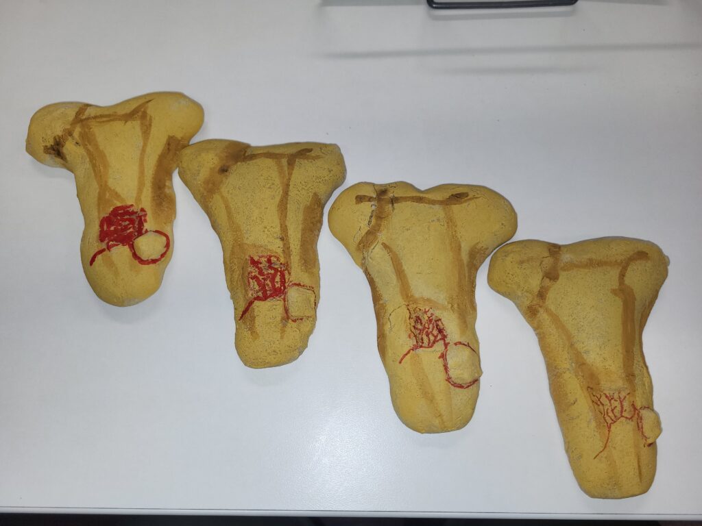
The Hauser procedure introduced by Emil Hauser in 1938 was the original procedure for for posteromedialization of the tibial tubercle. The purpose of the Hauser procedure was specifically to stabilize the patella by realigning the patellar tendon medially and pull the kneecap into a more stable position. The procedure would involve the surgeon cutting the tibial tubercle from the tibia, relocating it’s position medially, and securing the tubercle with screws or K wires. The procedure increases the tension on the patellar tendon and decreases the likelihood of repeated dislocation.
The Hauser procedure was common and long considered effective, for long term positive results, for the majority of the patients who had the procedure performed. However, it was not without complications. In addition to normal surgical complications there was the possibility of developing compartment syndrome or atrophy from extended recovery time. Normal recovery 3-4 weeks on crutches and in a cast or immobilizer, followed by more time in an immobilizer and physical therapy for generally 3-6 months.
In one study on the Hauser procedure, “The operation was considered to be successful in 52 of 62 cases. However, 36 knees caused pain or discomfort; in 2 cases the pain was severe. Patellar stability was achieved better in adults than in children. Only one fourth of the knees had both complete freedom from pain and stability”. Barbari, S et al., (1990).
“In 1983, Fulkerson introduced the anteromedialization technique and, later, straight anteriorization without bone grafts”. Saper, M et al., (2017). The procedure developed by Fulkerson moves the tibial tubercle both anterior and medial and is much better. Saper, M et al., (2017).
With the Fulkerson procedure the tibial tubercle is moved medially just slightly to avoid over correcting the alignment of the patellar tendon. Repositioning the patellar tendon changes the angle of the patella into a better position in line with the femur. The tibial tubercle is cut completely free from the tibia allowing it’s placement to be located in the best location for alignment whether that is anterior, medially, and distally or just medially. The tubercle is held in position with K-wires or screws as it is with the Hauser procedure and any bony defect is filled with bone putty.
Patients are released with a knee immobilizer and crutches and are partial weight for the first 6 weeks. Physical Therapy most often lasts only 4-6 weeks and the patient has regained strength in their quadriceps they can stop using the immobilizer. After the bone has healed effectively the patient can be cleared to return to regular activities.
Patients who had Fulkerson’s anteromedialization performed had a higher success rate. “86% (36 of 42) of the patients reported good or excellent results. Two reported fair results, and 4 had poor results”. Buuck, D et al., (2000).
Both procedures had comparable bone healing with blood clotting only hours after the cutting of the bone. Approximately 48 hours later chondrocytes create an internal callus between the pieces of bone. During that same time period chondrocytes and osteoblasts help stabilizable the bone by creating an external callus of hyaline cartilage and bone around the outside of the bone. Over the following weeks dead bone is reabsorbed by osteoclasts and osteoblasts are created. Ossification then replaces the calli with bone. When the internal and external calli meet spongy bone is replaced with compact bone and healing is complete.
The only noticeable difference is with the anteromedialization is that any bone deficit left by the removal and placement of the tibial tubercle is filled in with bone putty to assist in the healing of the bone by creating and artificial bone graft.
 Posted by
Bone Healing After a Hauser Procedure Compared to an Anteromedialization
The Hauser Procedure first introduced in 1938 was used as the primary surgical option for correction of recurrent traumatic dislocation of the patella for nearly half a century. In 1983 the first Fulkerson Osteotomy was performed leading the way to the beginning of the end for the Hauser Procedure.
The Hauser procedure introduced by Emil Hauser in 1938 was the original procedure for for posteromedialization of the tibial tubercle. The purpose of the Hauser procedure was specifically to stabilize the patella by realigning the patellar tendon medially and pull the kneecap into a more stable position. The procedure would involve the surgeon cutting the tibial tubercle from the tibia, relocating it’s position medially, and securing the tubercle with screws or K wires. The procedure increases the tension on the patellar tendon and decreases the likelihood of repeated dislocation.
The Hauser procedure was common and long considered effective, for long term positive results, for the majority of the patients who had the procedure performed. However, it was not without complications. In addition to normal surgical complications there was the possibility of developing compartment syndrome or atrophy from extended recovery time. Normal recovery 3-4 weeks on crutches and in a cast or immobilizer, followed by more time in an immobilizer and physical therapy for generally 3-6 months.
In one study on the Hauser procedure, “The operation was considered to be successful in 52 of 62 cases. However, 36 knees caused pain or discomfort; in 2 cases the pain was severe. Patellar stability was achieved better in adults than in children. Only one fourth of the knees had both complete freedom from pain and stability”. Barbari, S et al., (1990).
“In 1983, Fulkerson introduced the anteromedialization technique and, later, straight anteriorization without bone grafts”. Saper, M et al., (2017). The procedure developed by Fulkerson moves the tibial tubercle both anterior and medial and is much better. Saper, M et al., (2017).
With the Fulkerson procedure the tibial tubercle is moved medially just slightly to avoid over correcting the alignment of the patellar tendon. Repositioning the patellar tendon changes the angle of the patella into a better position in line with the femur. The tibial tubercle is cut completely free from the tibia allowing it’s placement to be located in the best location for alignment whether that is anterior, medially, and distally or just medially. The tubercle is held in position with K-wires or screws as it is with the Hauser procedure and any bony defect is filled with bone putty.
Patients are released with a knee immobilizer and crutches and are partial weight for the first 6 weeks. Physical Therapy most often lasts only 4-6 weeks and the patient has regained strength in their quadriceps they can stop using the immobilizer. After the bone has healed effectively the patient can be cleared to return to regular activities.
Patients who had Fulkerson’s anteromedialization performed had a higher success rate. “86% (36 of 42) of the patients reported good or excellent results. Two reported fair results, and 4 had poor results”. Buuck, D et al., (2000).
Both procedures had comparable bone healing with blood clotting only hours after the cutting of the bone. Approximately 48 hours later chondrocytes create an internal callus between the pieces of bone. During that same time period chondrocytes and osteoblasts help stabilizable the bone by creating an external callus of hyaline cartilage and bone around the outside of the bone. Over the following weeks dead bone is reabsorbed by osteoclasts and osteoblasts are created. Ossification then replaces the calli with bone. When the internal and external calli meet spongy bone is replaced with compact bone and healing is complete.
The only noticeable difference is with the anteromedialization is that any bone deficit left by the removal and placement of the tibial tubercle is filled in with bone putty to assist in the healing of the bone by creating and artificial bone graft.
Barbari, S., Raugstad, T.S., Lichtenberg, N., Refvem, D. (1990). The Hauser operation for patellar dislocation. Acta Orthopaedica Scandinavica, 61(1), 32-35. https://www.tandfonline.com/doi/epdf/10.3109/17453679008993061?needAccess=true
Buuck, D.A., Fulkerson, J.P. (2000). Anteromedialization of the Tibial Tubercle: A 4- to 12- year follow-up. Operative Techniques in Sports Medicines, 8(2), 131-137. https://onlinebiol111fall2025.slack.com/archives/D09E50NJCQM/p1763059412630939
Saper, M.G., Cox, B.A., Shneider, D.A. (2017). A modified osteotomy for anteromedialization of the Tibial Tubercle. Arthroscopy Techniques, 6(4), e1107-e1112. https://www.sciencedirect.com/science/article/pii/S2212628717300889e
Betts, J.G., Desaix, P., Johnson, E., Johnson, J.E., Korol, O., Kruse, D., Poe, B., Wise, J.A., Womble, M., Young, K.A. (2022). Anatomy and Physiology (2, 214-215). Openstax.