 Posted by
I chose to connect my STEAM project to unit 4 objective “Know the structure of the knee joint and compare this joint to the other 5 joint types”
The achilles tendon is the longest tendon in the body. It is approximately 6 centimeters long. It is very susceptible to acute and chronic injury due to its size. (DeMaio et al., 1995) It is a thick rope-like structure. It attaches the calf muscles to the foot. (Marx, 2005) It is a continuation of the triceps surae. The triceps surae are made up of the muscles heads gastrocnemius and soleus. It has an origin from medial to lateral femoral condyles. It inserts into the middle third of the posterior tuberosity of the calcaneus. (Myerson and McGarvey, 1998)
There is a connective tissue called peritenon. It varies in thickness and surrounds the tendon. It is also called paratenon. The tendon itself is made out of collagen fibers. It gets the fibers from the gastrocnemius and soleus muscles. (DeMaio et al., 1995)
Achilles tendonitis is inflammation of the achilles tendon. The most common cause is due to overuse of the tendon. It also happens when the calf muscles gastrocnemius and soleus are tight. (Marx, 2005) It is most common in athletes that run, jump, and impact the lower extremities. It is also from repetitive motions and force on the tendon beyond its ability to heal.(Myerson and McGarvey, 1998) During running the tendon is subjected to 4 to 6 times our body weight. During walking it is subjected to 2 to 3 times our body weight.(DeMaio et al., 1995) The tendon is both vascular and avascular. It is avascular 3 to 5 centimeters proximal to insertion. The avascular area is prone to chronic tendinitis. (Myerson and McGarvey, 1998) It is also more likely to happen as we age. This is due to degeneration of the tendon and vascular supply. It can also be the shoes and surface that is being run on. (DeMaio et al., 1995)
The symptoms are pain and swelling during and after physical activities. Pain and stiffness in the morning. This is because people usually sleep with their toes pointed. Tenderness and warmth to the touch. (Marx, 2005) There is acute, subacute, and chronic tendonitis. When it is chronic it also becomes weaker. (DeMaio et al., 1995)
Achilles tendonitis can be classified into three subcategories. paratendinitis, tendinosis, and paratendinitis with tendinosis. Although diagnostic imagery is rarely needed. They found these categories with tissue biopsies and ultrasound and magnetic resonance imaging.(DeMaio et al., 1995) Now they diagnose it by putting pressure on the tendon with their index finger and thump. (Myerson and McGarvey, 1998)
Paratendinitis is only inflammation of the tendon lining, not the tendon itself. When diagnosing they use the finger method. During the examination thickness and tenderness is mentioned. They also noted a crackling or popping sound when skin is glided over the tendon. Tendinosis is inflammation of the tendon, not the peritenon. It is also degeneration of the tendon due to age, microtrauma and decreased vascular supply. There are also inflammatory cells found in the area of degeneration. The tendon also appears soft, yellow, and dull looking. For paratendinitis with tendinosis, both the tendon and peritenon are inflamed. Before diagnosing they noticed visible thickness. During examination there is pain when squeezed. Tendon is also softer and more yellow. Acute symptoms usually last two weeks. Subacute symptoms usually last three to six weeks. Chronic can last six weeks and longer. If symptoms last more than six months with treatment surgery may be recommended. (DeMaio et al., 1995)
The treatment recommended is rest and stretching. They need to change their activity level while doing stretching exercises. If it continues a boot may be necessary. They also recommend non-steroidal anti-inflammatory medication. (Marx, 2005)
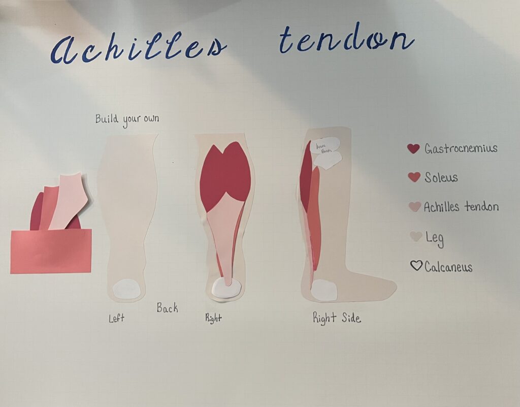
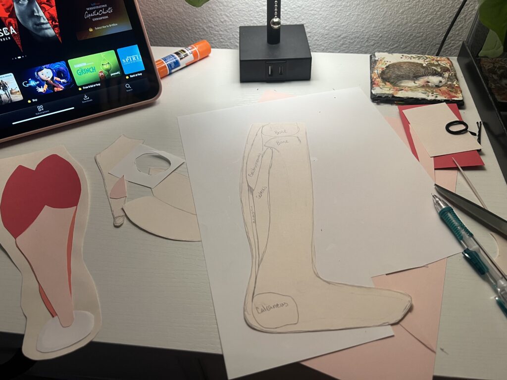
For my abstract art to go with my essay I chose to use construction paper. I used visible body suite as a reference while drawing the achilles tendon. Along with the muscles gastrocnemius and soleus.(Argosy Publishing, n.d.). I was originally going to just draw the tendon and muscles. I decided to go beyond that because I wanted to add texture and feel to it. I layered the construction paper on top of each other to create it. I also used different colors for the different muscles and the tendon. I added a color key so they are easily identifiable. I made two different points of views. One from the back and one from the side. I chose to do two different views because the soleus muscle has a better view from the side. For the view from the back I glued the right side down and not the left side. I chose to do this because I wanted it to be able to be taken apart.
Work cited
Argosy Publishing (n.d.) Achilles tendon (calcaneal tendon, triceps surae, part) inflames, R. Retrieved November 19, 2025, from https://apps.visiblebody.com/share/?p=vbhaa&t=4_310595_638993493148360000_154119
DeMaio, Marlene,M.C., U.S.N.R., Paine, R., P.T., & Drez, David J,Jr, M.D. (1995). ACHILLES TENDONITIS. Orthopedics, 18(2), 195-204. Retrieved from https://www.proquest.com/scholarly-journals/achilles-tendonitis/docview/962455009/se-2
Marx, R. (2005, 05). ACHILLES TENDINITIS. Joe Weider’s Muscle & Fitness, 66, 192. Retrieved from https://www.proquest.com/magazines/achilles-tendinitis/docview/216367585/se-2
Myerson, M. S., & McGarvey, W. (1998). Disorders of the insertion of the achilles tendon and achilles tendinitis. Journal of Bone and Joint Surgery, 80(12), 1814. Retrieved from https://www.proquest.com/scholarly-journals/disorders-insertion-achilles-tendon-tendinitis/docview/205146104/se-2
Charlisa did her STEAM project on Achilles Tendonitis. The Achilles tendon is the bodies longest tendon. It is put under an incredible amount of stress when we run and walk. The avascualr section 3-5 inches above the heel is especially vulnerable. Achilles Tendonitis is an inflammatory injury in this tendon, often caused by overuse, tightness in that area or the process of natural ageing. It normally starts in this avascular region above the heel. Symptoms include swelling, morning stiffness, tenderness, and warmth in the area. This condition can be acute, subcute, or chronic. Chronic being the most severe, and acute being the least severe. In the past health care professionals used different imaging techniques to diagnose this condition. Now, a diagnostic is as simple as a physical examination and palpation of the ankle. Less severe cases go away after a few weeks. If symptoms persist over long periods of time, surgery may be recommended. For less severe cases, over the counter pain medication, decreased activity, rest and special stretches can suffice.
Charlisa’s abstract is a build your own Achilles tendon poster, made with construction paper. She layered her paper to create a textured effect.
I didn’t know anything about this condition before this STEAM project, and I may refer a friend who is dealing with similar symptoms to this short essay. It was an efficient, concise overview of the condition, and written in a way the general population can understand.