 Posted by
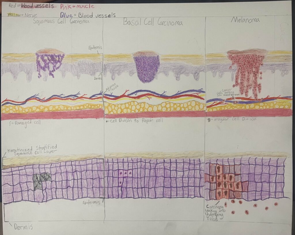
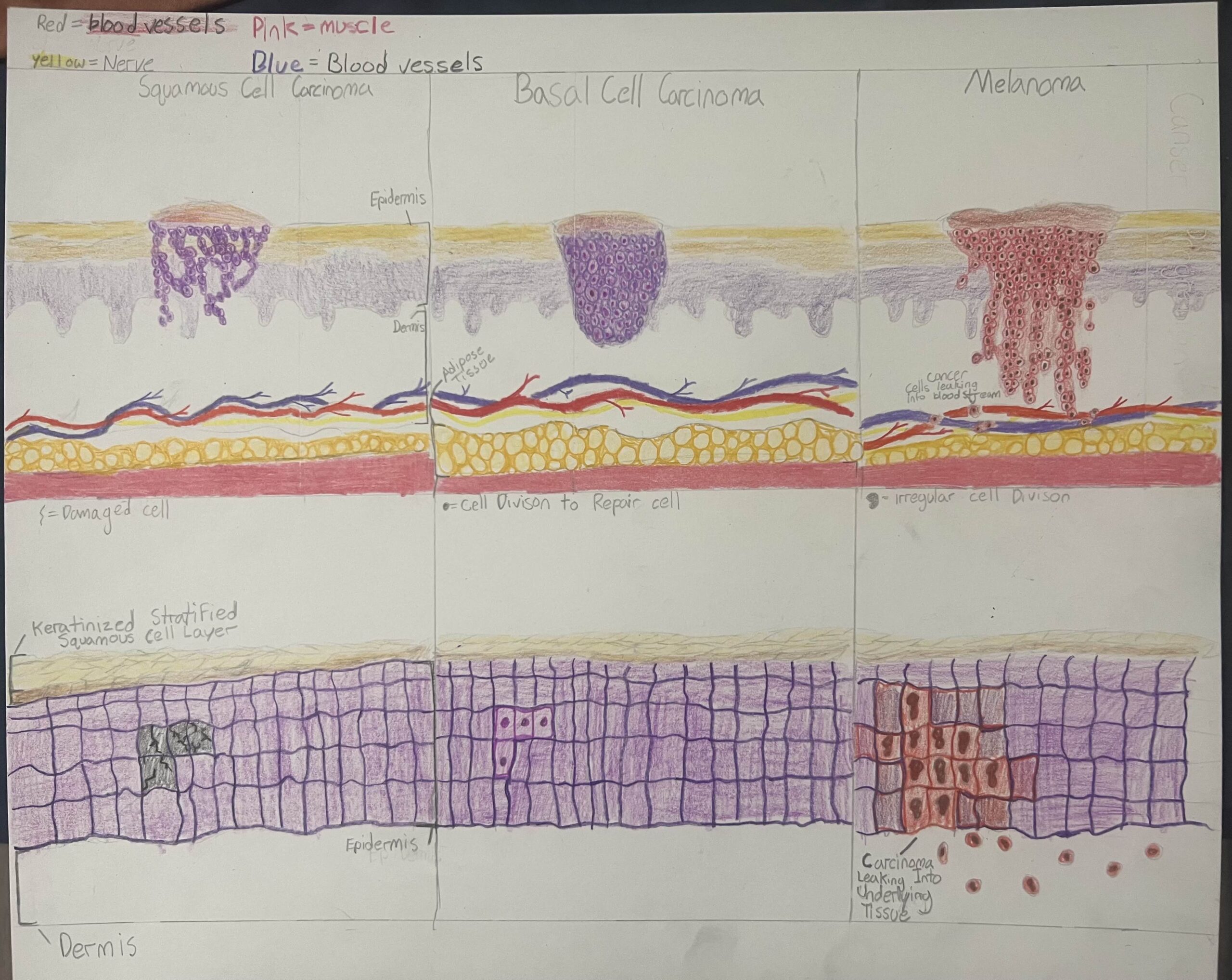
Skin is the first line of defense in the body. The skin is made up of a type of epithelial tissue known as stratified squamous epithelium. The stratified squamous epithelium is stacked layers of scale like cells. The skin surface layer is made up of a specific type of stratified squamous epithelium known as keratinized stratified squamous epithelium (Betts et al., 2021). The surface skin cells are squamous epithelial, whereas the basal (deeper layers of skin) contains either columnar or cuboidal squamous epithelial cells (Betts et al., 2021). These layers of skin are put through a lot of stress such as tearing, burns, and much more. Because they are put through so much, they are constantly repairing. The steps of repair are as follows. First is inflammation. This is caused by cell death and limits further injury, partially or fully eliminates the foreign object causing injury, and starts the repair and regeneration of injured tissue (Betts et al., 2021). Next, blood starts filling the wound bringing white blood cells to help remove dangerous foreign material and that same blood hardens to prevent further infectious material from entering. After that, the epithelial tissues can start to repair. Epithelial tissues repair through a process known as mitosis. Mitosis has three stages that are triggered by cyclin-dependent kinase 1 (Crncec & Hochegger, 2019). Prophase is the first phase of mitosis and is when chromosomes condense and become visible (Betts et al., 2021). Next phase, metaphase, the chromosomes are lined up at the metaphase plate of the cell (Betts et al., 2021). After this, during anaphase, the chromosomes split in two (Betts et al., 2021). Then, during telophase, the chromosomes at opposite poles begin to decondense and the nuclear envelope surrounds them (Betts et al., 2021). Lastly, they split apart in a phase known as telophase (Betts et al., 2021). These tissue cells can become cancerous. Cancerous cells form when the DNA is mutated during mitosis and the cells continue to multiply at an uncontrolled rate (Hasan et al., 2023). There are different types of skin cancer; squamous cell carcinoma, basal cell carcinoma, and melanoma (Skin Cancer – Symptoms and Causes, n.d.). Squamous cell is the second most common type of skin cancer. It is typically generalized to the epidermis and looks like scaly patches of red firm bumps. Basal cell carcinomas are the most common type of cancer. They form and reach down from the epidermis to the dermis of the skin. It is not known to be that aggressive and has a low mortality rate. Though it is not known to be aggressive, it can spread through the surface of the body and can escalate to bone and nerves (Hasan et al., 2023). The basal cell carcinomas look like pinkish bumps on the skin. Melanoma is the most dangerous out of all types of skin cancer. It goes deeper than the epidermis and dermis, reaching all the way down to the blood vessels. This is dangerous because the cancerous cells of a melanoma get into the blood stream, spreading and interrupting the functions of organs, which can make it deadly if not treated (Hasan et al., 2023). The way they form is from melanocytes. Melanocytes are located in the epidermis. The reason melanomas form is when a buildup of genetic alterations happens, deactivating tumor suppressor genes and stopping DNA repair occur (Hasan et al., 2023). This could result in accelerated melanocyte growth causing melanomas. Melanomas are characterized by a brownish red color and their diameter is greater than 6 millimeters. There are many ways you can get skin cancer. The main way you can get skin cancer is through UV rays. UV rays damage the cells, then induce cell death (adiposis), and hinder DNA repair, which can cause mutations in DNA (Hasan et al., 2023). UV rays can also cause the deconstruction of DNA, which can cause damage and cause mutations that can lead to skin cancer. That is why tanning beds and sun burns are terrible for your skin (Skin Cancer – Symptoms and Causes, n.d.).
In T-Rex’s project, the diagram of skin cells helped me understand the differences between healthy and cancerous skin cells. Right off the bat, he explains the skin’s purpose. It is there to protect the body and serve as the first line of defence. The skin’s first layer starts with the epidermis, made of primarily stratified squamous cells. Deeper into the epidermis layer, the basal layer is made up of either cuboidal or columnar epithelial cells. When the skin gets damaged, there are steps for repair and regeneration—starting with inflammation to prevent further cell death and ending with blood clots. Afterward, epithelial tissue can regenerate through mitosis. Then, T-Rex explains how mitosis can be disrupted when skin cancer develops.
The most common way to get skin cancer is through UV rays, such as sunburn and tanning beds, because they damage the epithelial cells and cause DNA mutations. The mutation affects mitosis, causing the cells to multiply at an uncontrolled rate. Cancerous cells can appear in the squamous or basal layers or in melanocytes. Basal cell carcinoma is the most common, followed by squamous cell carcinoma. They are known for being nonaggressive. They can be seen on the skin’s surface and can spread to bones and nerves if left untreated. Melanoma arises from melanocytes in the epidermis and is the most aggressive skin cancer, metastasizing to other organs. Melanoma changes the genetics, which might cause tumors to appear. T-Rex diagram shows how each type of cancer spreads.