 Posted by
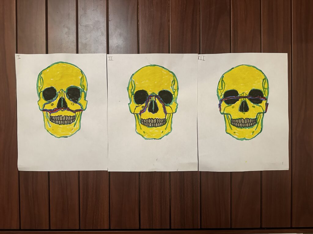
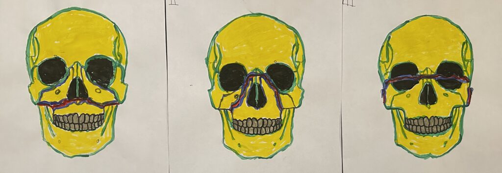
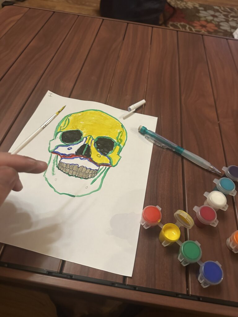

My kids played rock, paper, scissors to decide who got to choose the colors. My daughter won and picked yellow and green. The red indicates the fracture line, and while the blue didn’t really come out well, it’s supposed to indicate where osteoblasts would begin repairing the bone.
I used skull outlines provided by wikipedia user RosarioVanThulpe. Le Fort 1 Fracture (File:LeFort1a.png) by RosarioVanTulpe / Wikimedia Commons, used under CC BY-SA 3.0 (and GNU FDL) – adapted for this art project.
Abstract of “Le Fort Fracture Classifications” by Trevin Lovett
In this STEAM project, Lovett creates three diagrams, displaying the three different Le Fort fracture types. In these diagrams, red indicates the location of the fracture, while blue indicates where osteoblasts begin to repair the bone structure. The first fracture indicated is the, “Le Fort type I fracture,” which separates the maxilla from the midface. This type of fracture is the result of low-energy blows to the upper mouth region. The second is, “Le Fort type II fracture,” which is a pyramidal shaped fracture that impacts the maxilla, nasal bridge, lacrimal bones, and infraorbital rims. This second pattern results from higher-energy impacts to the central midface. The last fracture shown is the, “Le Fort type III fracture,” a craniofacial dissociation separating the midface from the cranial base. This third pattern is the most severe, resulting from high-energy trauma.
Le Fort fracture types encompass a system of identifiable, high-impact fractures that center around structures of the midface. This fracture identification system is critical for treatments, as the location of these injuries can lead to immediate threats to airways, speech, and nutrition. This system allows for the rapid communication between EMTs, nurses, and physicians.
This project satisfies the learning objective of classification of bone fractures from unit four of the course.