 Posted by
Bone development is a complex and highly coordinated process that depends on cellular activity, structural proteins, and the endocrine system. For this STEAM project, I focused specifically on two course objectives, how bone development is hormonally regulated and the stages of bone development and repair. To explore these concepts, I examined Osteogenesis Imperfecta (OI) in adolescence, “a genetic disorder that is characterized by increased bone fragility and low bone mass” (Rauch & Glorieux, 2006) that disrupts both normal bone formation and the body’s ability to maintain a healthy skeletal structure.
During normal growth, hormones such as growth hormone, thyroid hormone, estrogen, and testosterone regulate osteoblast and osteoclast activity. They influence how quickly bones lengthen, how they remodel, and how efficiently they repair daily micro-damage. These hormonal signals depend heavily on the presence of strong connective tissue, especially Type I collagen, which forms the framework that minerals attach to during bone formation (Martin & Shapiro, 2007). In OI, “mutations affecting four genes, most often diverse mutations affecting type 1 collagen genes” (Martin & Shapiro, 2007) make the collagen framework defective. This means that even when hormonal regulation is functioning normally, bone development remains impaired. Completing this project helped me understand how hormones and bone matrix structure interact, and how one malfunction can disrupt the entire developmental process.
We also learned the stages of bone development and repair. Most children’s bone growth occurs through endochondral ossification, where a long bone first forms as a hyaline cartilage model (Betts, 2022). Mesenchymal cells become chondrocytes, the cartilage enlarges, a calcified matrix forms, and capillaries invade to create the primary ossification center (Betts, 2022). Later, secondary ossification centers develop in the epiphyses, leaving the epiphyseal plate as the site of continued growth. In OI, these steps still occur, but the collagen-based framework supporting these processes is defective from the beginning. Even when cartilage is replaced by bone, the resulting tissue is poorly organized and insufficiently mineralized, making fractures common.
Normally, a fracture triggers a hematoma, followed by internal and external callus formation made of fibrocartilage and early bone. These steps represent the stages of hematoma formation, soft callus development, hard callus formation, and finally bone remodeling (Marsell & Einhorn, 2011). Over time, the callus is replaced through endochondral ossification as trabecular bone forms, and later the region is remodeled into compact bone (Betts, 2022). In OI, the body attempts to follow the same repair sequence, but the abnormal collagen results in a weak callus and fragile new bone (Betts, 2022). Even after remodeling, the healed bone may remain structurally compromised. Seeing how OI alters both formation and repair helped me better understand why healthy collagen is essential at every stage.
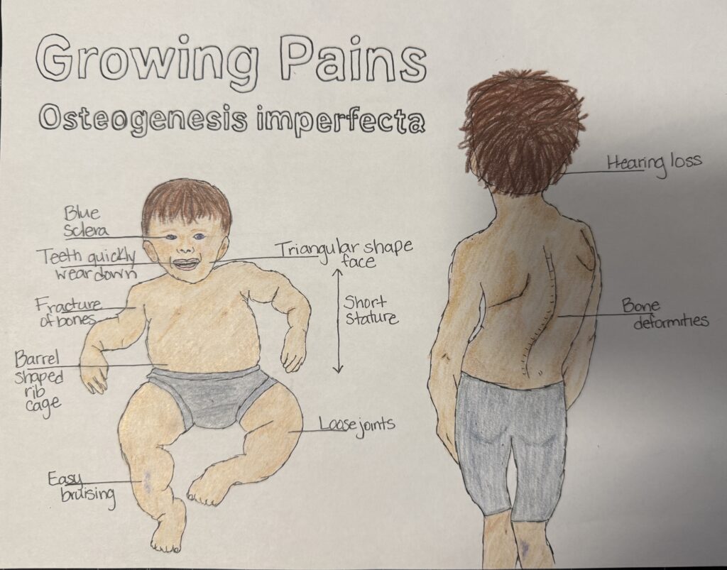
My artwork depicts two stages of childhood to show how OI affects development over time. The infant illustration highlights severe features such as “blue sclera, hearing loss, and short stature” (Martin & Shapiro, 2007). These symptoms reflect weakened connective tissue throughout the body, not just the skeleton. I also included loose joints, triangular facial structure, and high fracture susceptibility, effects linked to the inability to form stable, mineralized bone without strong collagen. The older child in my drawing shows long-term consequences including scoliosis, bowed legs, short stature, and potential hearing loss from abnormalities in the tiny ear bones (Martin & Shapiro, 2007). Presenting symptoms across two ages allowed me to demonstrate how the disorder interacts with natural developmental milestones from infancy through adolescence.
Overall, this project helped my understanding of how genetics, hormones, and structural proteins contribute to healthy bone development. Through my artwork, I was able to connect the course objectives, hormonal regulation of bone development and the stages of bone formation and healing, with a visual demonstration of how these processes fail in individuals with OI. Completing this STEAM project allowed me to combine creativity with scientific accuracy and strengthened my understanding of human development.
References
Betts, G. J. (2022, April 20). 6.4 Bone Formation and Development – Anatomy and Physiology 2e | OpenStax. Openstax.org. https://openstax.org/books/anatomy-and-physiology-2e/pages/6-4-bone-formation-and-development
Marsell, R., & Einhorn, T. A. (2011). The Biology of Fracture Healing. Injury, 42(6), 551–555. https://doi.org/10.1016/j.injury.2011.03.031
Martin, E., & Shapiro, J. R. (2007). Osteogenesis imperfecta: Epidemiology and pathophysiology. Current Osteoporosis Reports, 5(3), 91–97. https://doi.org/10.1007/s11914-007-0023-z
Rauch, F., & Glorieux, F. H. (2006). Treatment of children with osteogenesis imperfecta. Current Osteoporosis Reports, 4(4), 159–164. https://doi.org/10.1007/s11914-996-0025-2
Abstract:
This drawing is amazing first of all, (and the paper,) but in summary, Osteogenesis Imperfecta is a bone disease that leads to pains and fragility. This project explores how Osteogenesis Imperfecta (OI) affects childhood skeletal development by integrating two core course objectives: hormonal regulation of bone growth and the stages of bone formation and repair. The artwork depicts both an infant and an older child to highlight how symptoms of OI progress across developmental milestones. In the infant, features such as blue sclera, loose joints, a barrel-shaped rib cage, triangular facial structure, rapid tooth wear, and frequent fractures visually represent the widespread consequences of defective type I collagen. In the older child, scoliosis, bowed legs, short stature, and hearing loss further illustrate how repeated fractures, weak bone matrix structure, and impaired remodeling accumulate over time.
The written portion explains how growth hormone, thyroid hormone, estrogen, and testosterone normally guide bone lengthening, density, and continuous remodeling. However, in OI, hormonal signals cannot properly support bone development because the collagen scaffold that minerals attach to is structurally compromised from the start. The project also examines how OI alters major steps of endochondral ossification, beginning with cartilage formation and extending through primary and secondary ossification centers, resulting in bone that is insufficiently mineralized and easily deformed. In addition, the stages of fracture healing, including hematoma formation, soft and hard callus development, and bone remodeling, are addressed to show why repair in OI produces fragile, disorganized bone even after the healing process is complete.
Overall, this project demonstrates how genetics, hormones, and structural proteins collectively shape bone development, and how a single defect in collagen profoundly influences childhood growth and skeletal integrity.