 Posted by
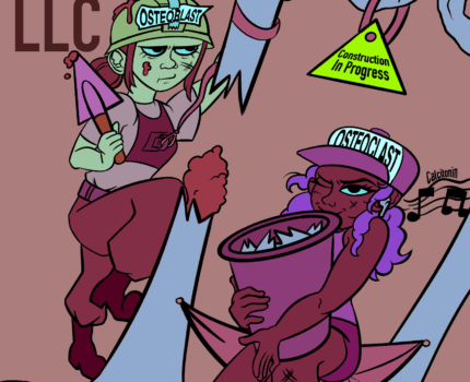
Here is my submission for Professor Don’s BIOF111X Fall course. I completed this entirely in Procreate. Proof of authenticity is provided below. Sorry if this platform slightly affects the render quality of the exported piece.
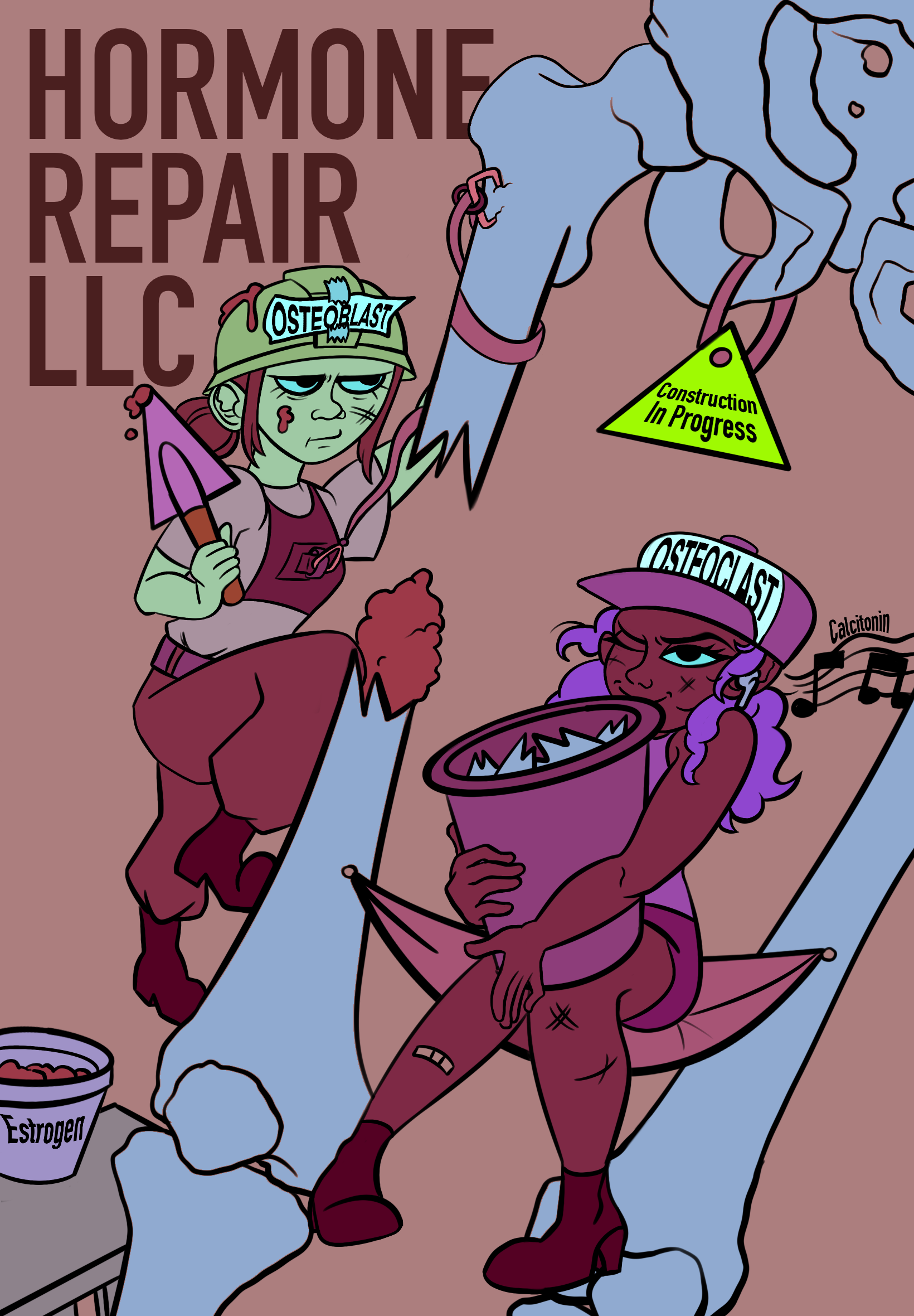
My STEAM project, called Osteoblasts & Osteoclasts At Work, uses digital images to show how bones grow, heal, and are controlled by hormones. Rather than just listing cell types, it shows osteoblasts and osteoclasts as specialized laborers repairing a fractured bone, mirroring real recovery stages. Visual storytelling highlights cooperation among these cells under hormonal influence; one forms new tissue, whereas the other removes injured tissue, underlining the key equilibrium needed for effective repair. Each phase illustrates that mending takes time through the synchronized actions of biological functions and chemical messengers.
This project supports several learning aims. For instance, it clearly shows how hormones control bone development and healing, aligning with course outlines on hormone functions in maintaining bone health. Important hormones like calcitonin and estrogen are presented as central controllers of cell behavior: calcitonin is illustrated as reducing osteoclast activity, mirroring its function in limiting bone breakdown; at the same time, estrogen is shown close by, working to maintain structural integrity. Instead of using detailed text, the image relies on clear visuals, making it easier for learners to grasp intricate biological processes.
Much like a diagram, the image helps illustrate four central bone cell types: osteoblasts, osteoclasts, osteocytes, and lining cells. Rather than just naming them, it shows osteoblasts and osteoclasts moving, which underlines their tasks – forming new bone or breaking down old parts, mainly when bones heal. Because they’re drawn in active positions, viewers grasp better what each does and how they work together. Instead of listing facts, the drawing suggests broader duties of bone, including giving shape, protecting vital areas, holding key minerals, and connecting those roles to fixing breaks.
Basing this study on up-to-date science makes it more useful for teaching. Bone repair occurs in four key stages: swelling initiates it, followed by soft-tissue buildup, then rigid callus formation, and finally reshaping (Einhorn & Gerstenfeld, 2015). At first, bone-eating cells clear out damaged areas; at the same time, bone-building cells lay down fresh material that gradually becomes stronger. Hormones guide these steps – calcitonin slows breakdown, but when blood calcium drops, parathyroid hormone speeds it up. Estrogen helps bone-building cells function properly while preventing excessive breakdown, thereby preserving skeletal strength (Florencio-Silva et al., 2015). This initiative illustrates those intricate mechanisms using a building-site metaphor, turning chemical reactions into an easier-to-follow story.
The piece highlights how hormones are key to repairing and maintaining bone health. While calcitonin reduces osteoclast activity, PTH boosts it, showing that hormone signals guide cell behavior. Although estrogen supports osteoblast function, it also limits breakdown, underscoring the delicate hormonal balance needed for strong bones.
Overall, Osteoblasts & Osteoclasts At Work shows how visuals can boost learning. Instead of just text, it uses digital imagery grounded in science to explain bone recovery and hormone control. Rather than simply listing facts, the project hits core teaching goals while sparking curiosity about the body’s hidden repair methods. In fact, it highlights how cells, hormones, and minerals work together, linking different systems to repair tissues and support strong bones. Thanks to these visual narratives, I have grown to grasp complicated healing steps more easily, seeing them not as isolated events but as coordinated actions across the body. Healing turns out to be active, layered, yet surprisingly organized when shown through such clear images.
Einhorn, T. A., & Gerstenfeld, L. C. (2015). Fracture healing: Mechanisms and interventions. Nature Reviews Rheumatology, 11(1), 45–54. https://doi.org/10.1038/nrrheum.2014.164
Florencio-Silva, R., Sasso, G. R. S., Sasso-Cerri, E., Simões, M. J., & Cerri, P. S. (2015). Biology of bone tissue: Structure, function, and factors that influence bone cells. BioMed Research International, 2015, 421746. https://doi.org/10.1155/2015/421746
Grammarly. (2024). Grammarly Handbook.
Martini, F. H., Nath, J. L., & Bartholomew, E. F. (2021). Fundamentals of anatomy & physiology (12th ed.). Pearson Education.