 Posted by

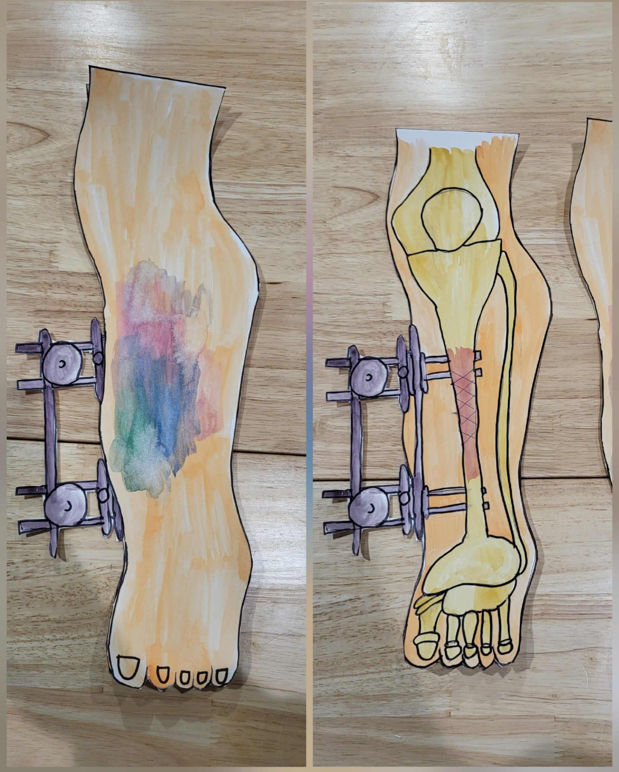
The visual provided displays first a leg with bruising on the outside indicating an injury. The item to the left is what an external fixation device looks like from the outside, it is a collection of metal plates and pins/wires used to help stabilize the leg. The second photo is what this device looks like below the skin and what exactly is happening to the bone. The external fixation device has prongs that extend to either side of the fractured bone and hold it in place careful to not further irritate the impacted area. While this type of surgery seems new, the concept of supporting bones externally has been applied for over 2000 years (Bliven et al., 2019).
External fixation surgery a useful and life saving medical device and procedure, this essay and projects aims to explore more about this process; the course objective covered will be classifying fractures and the stages of bone development and repair. When an individual breaks or fractures their bone and requires surgery to fix it, there are many different kinds of procedures and ways to mend the fracture and help it, help the patient heal quickly and feel better faster. Common surgical tools include casts, and braces, but for complicated tibial fractures, external fixation is one of the most utilized treatment options (Albushtra et al., 2024). In its essence, external fixation is a procedure that entails the use of percutaneously (through the skin) implanted transosseous (through the bone) pins and wires which are secured to external scaffolding in order to provide support and protection for a limb. Using this method ensures that the bone or joint can be stabilized. This method of surgery needs to meet three conditions to allow success: the pins and wires need to avoid damage to vital structures, allow easy access to the area of injury, and should meet the mechanical demands of the patient and the injury (Fragomen et al., 2006).
Femoral and tibial fractures are classified using a set list produced by the Orthopaedic Trauma Association, the system gives each fracture an alphanumeric code which allows easier classification and recognition based on anatomical location, fracture morphology, and complexity. While other classifications do exist, this one is the easiest and most available: A “Type 1” fracture is a minimal soft tissue injury, such as a simple fracture where the wound is no more than 1 centimeter. A “type ii” would be an injury where soft tissue coverage is adequate, there is minimal comminution (fracture) but the wound is greater than 1 cm. A “type iiiA” is a high energy injury, there is extensive soft tissue damage, the fracture is contaminated and comminuted, however the bone remains adequately covered. A “Type iiiB” is a high-energy injury, there is extensive soft tissue damage and periosteal stripping and the bone is exposed. Lastly, a “type iiiC” is similar to iiiB but also has an arterial deficit that needs repairing. Usually the more severe injuries require external fixation surgery (Yates, et al., 2016). The tibia is one of the most fractured long bones, partly due to its location which exposes it to a higher incidence of trauma, the most common being road traffic accidents. These injuries are considered extreme due to additional soft tissue damage or damage to the neurovascular bundle. Additionally, there are many other risks with this type of injury, including malunion, non-union, infections, or a general need for surgical re-intervention (Albushtra, et al., 2024).
Bone has a rare capability to regenerate without scars, in fact it is able to fully restore the injured bone areas, and new technologies are developing to assist in bone injury recover, only approximately five to ten percent of all bone fractures fail to heal or heal in a delayed way, however this usually involved various risk factors among patients and the specifics of their injuries (Steppe, et al., 2023). Bone fracture healing involves cellular and molecular components. There are two types of bone healing: the first is direct, where healing entails a direct shift of mesenchymal stem cells to osteoblasts that form bones. The second is indirect, where the healing moves through a cartilage intermediate before a bone is created by osteoblasts. Most fractures clinically heal in the second method of healing, so that is what will be examined in this section. The first phase of bone healing is known as the inflammatory phase, which has three parts; acute inflammation, resolving inflammation,and chronic inflammation. During hemorrhage of the injured site, inflammatory cells are deposited along the clot. Some important functions of inflammatory cells is the removal of injured and devitalized tissue, as well as creating growth factors such as fibroblast growth factors, platelet derived growth factor, and growth factor beta, which begins the repair mechanisms. Despite the inflammatory stage being the earliest phase of repair, inflammatory cells are also found in later stages and change in various ways to assist healing. The next stage of bone repair is the fibrovascular phase, where the primary target is vascular remodeling. When the bone initially gets hurt, the vascular supply is remade by developing a new vascular network, this is done via angiogenesis and vasculogenesis. Angiogeneiosis is the process of new blood vessels being created, this is done by building off already present vasculature. Vasculogenesis is the new creation of blood vessels from progenitor cells. This process is extremely important in making sure bone tissue has blood and is kept alive. The next step in the healing process is bone formation. Bone formation is done with osteoblasts and chondrocytes. Osteoblasts bridge across the marrow cavity and build up the bone, while chondrocytes are highly angiogenic and create another run of vascular invasion into the cartilage. Together, the new bone is able to grow and heal. The last stage of bone healing and formation is called callus remodelling. In this last stage, osteoclasts are responsible for bone degradation, they remove the old and damaged bone which has been replaced by new bone via osteoblasts (Rodriguez-Merchan, 2024).
This visual was created to show what these types of fractures look like on the surface, but also the internal healing process and how tools can assist. While metal rods and bruises help with the outside, the most important processes are happening internally.
Lydia’s project explores external fixation surgery, a crucial medical procedure for treating complex tibial fractures. The work begins with a visual representation of the external fixation device, both externally and internally, highlighting its role in stabilizing fractured bones. The essay delves into the classification of femoral and tibial fractures using the Orthopedic Trauma Association’s alphanumeric system, emphasizing the severity levels that often require external fixation.
The project then examines the bone healing process, detailing its phases: inflammatory, fibrovascular, bone formation, and callus remodeling. It explains the cellular and molecular components involved in each stage, from the initial inflammatory response to the final remodeling of the bone structure. The work emphasizes the remarkable ability of bones to regenerate without scarring, noting that only a small percentage of fractures face healing complications.
Lydia’s research underscores the importance of external fixation in treating severe tibial fractures, particularly those resulting from high-energy traumas like road accidents. The project effectively combines visual elements with detailed explanations of surgical procedures and biological processes, providing a comprehensive overview of bone fracture treatment and healing mechanisms. This work demonstrates a thorough understanding of the course objectives related to fracture classification and bone development and repair.