 Posted by
For my STEAM project, I chose the topic of Supraventricular Tachycardia (SVT), which aligns with Course Objective #39: “How the heart translates an electrical signal into a contraction.”
The human heart relies on an intricate and consistent electrical conduction system to maintain its rhythmic contractions. Every heartbeat begins with an electrical signal that follows a specific pathway, initiating contractions that circulate blood throughout the body. SVT represents a significant deviation from this normal process and highlights the importance of understanding the heart’s electrical function.
The heart’s electrical system begins in the sinoatrial (SA) node, the heart’s intrinsic pacemaker. This property of initiating impulses spontaneously is called automaticity. From the SA node, the impulse travels through the atria, causing them to contract or squeeze pushing the blood into the ventricles. It then reaches the atrioventricular (AV) node, briefly delays to allow for ventricular filling, and continues through the bundle of His, bundle branches, and Purkinje fibers at the apex of the heart, where it initiates a powerful ventricular contraction (Nair et al., 2013). This well-orchestrated pathway ensures that the atria and ventricles beat in coordination, enabling efficient blood flow to the lungs and body.
This electrical activity can be measured using an electrocardiogram (ECG), which presents waves such as the P wave (atrial depolarization) and QRS complex (ventricular depolarization). A standard 12-lead ECG offers multiple views of the heart’s electrical activity, making it a valuable diagnostic tool for identifying arrhythmias, ischemia, and conduction abnormalities (Adeniran et al., 2013)
However, in some cases, the electrical impulse does not originate from the SA node. In Supraventricular Tachycardia, the signal may arise from a different, abnormal focus above the ventricles or may follow an alternative, reentrant pathway. This leads to rapid and often inefficient heartbeats, as both the atria and ventricles contract too quickly for adequate filling and ejection of blood. As a result, cardiac output is significantly reduced (American Heart Association, 2022).
SVT can be triggered by various factors such as dehydration, drug use, stress, or underlying cardiac disease. Symptoms of SVT include palpitations, dizziness, hypotension, shortness of breath, and in severe cases, altered mental status due to reduced cerebral perfusion (Kluger & Sweeney, 2009). If untreated, the condition can become life-threatening due to the heart’s inability to maintain effective circulation.
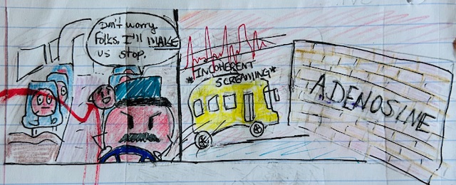
Fortunately, several treatments are available. The first line of treatment often involves stimulating the vagus nerve to slow conduction through the AV node. Techniques such as the Valsalva maneuver, facial immersion in ice water, or carotid sinus massage can activate the parasympathetic nervous system and help terminate the abnormal rhythm (Kluger & Sweeney, 2009). If vagal maneuvers are unsuccessful, adenosine, a rapid-acting medication, may be administered intravenously. Adenosine temporarily halts AV nodal conduction, giving the SA node a chance to reassert its control and restore normal sinus rhythm. While effective, this momentary cessation of the heartbeat can be distressing for both patients and providers.
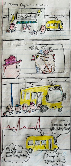
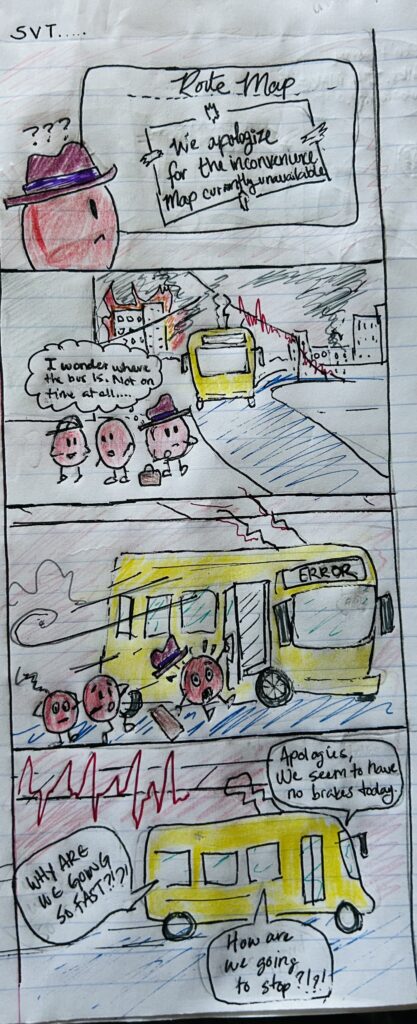
For my visual component, I created a comic strip that creatively illustrates the normal and abnormal pathways of cardiac conduction. In the story, red blood cells are personified as commuters traveling on a route through the heart, boarding a “bus” guided by the normal conduction system (SA → AV → Purkinje). As SVT begins, a rogue shortcut forms, and the heart starts firing too quickly. The bus speeds up, leaving many “passengers” behind, representing poor ventricular filling. Above the trolley, a power line displays the changing ECG waveform, transitioning from a normal sinus rhythm to a narrow-complex tachycardia. Finally, the “emergency brakes” are humorously applied when adenosine is administered, halting the chaotic rhythm and allowing the red blood cell commuters to resume their proper path.
This visual narrative emphasizes how even slight deviations in the electrical pathway can lead to serious physiological consequences. By incorporating imaginative elements with anatomical accuracy, the comic helps to illustrate the delicate balance required for effective cardiac function and how SVT disrupts that balance.
References
American Heart Association. (2022). Tachycardia – Fast heart rate. https://www.heart.org/en/health-topics/arrhythmia/about-arrhythmia/tachycardia–fast-heart-rate
Kluger, J., & Sweeney, M. (2009). Management of supraventricular tachycardia. Medical Journal of Australia, 190(5), 255–260. https://www.mja.com.au/system/files/issues/190_05_020309/med10727_fm.pdf
Adeniran, I., Hancox, J. C., & Zhang, H. (2013, December 17). Effect of cardiac ventricular mechanical contraction on the characteristics of the ECG a simulation study. SCIRP. https://www.scirp.org/journal/paperinformation?paperid=41405

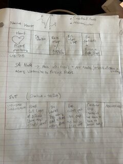
This was a very interesting piece on Supraventricular Tachycardia. The introduction was clear on the course objective. Within the paper, the course objective was covered nicely. The details about Supraventricular Tachycardia covered the definition of what it is, where it happens, and how to treat it. When reviewing this piece, I learned a few things that I feel went beyond what we learned in this class. For starters, I was unsure of what “His” bundles were, and I had to look them up. It was a new acronym I had not heard before to describe the atrioventricular bundle that extends from our AV nodes and talks to our Purkinje fibers. The second thing I was surprised to learn about the Valsalva maneuver. While it would be scary to have your heart rate irregular, things like putting your face in ice water or giving someone a carotid sinus massage to help regulate heartbeat are valuable information for anyone to know. The comic strip was a brilliant way to demonstrate how the Supraventricular Tachycardia works. The comic strip and the description clearly state what a normal path for our internal pacemaker is, and the abnormal route of the Supraventricular Tachycardia. This paper also included scientific peer-reviewed citations, including the American Heart Association, the Medical Journal of Australia, and the Journal of Biomedical Science and Engineering, to support the information presented. Overall, this was a well-written paper with new information on it and a wonderful art piece to accompany the paper for understanding. Nice job!