
 Posted by

Objective: Describe the structure and function of the heart.
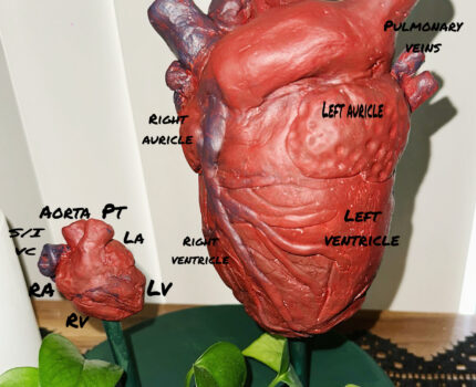
The human heart undergoes a remarkable transformation throughout our lifetime. In my STEAM project, I sought to show the actual size difference between the adult human heart and the newborn heart size (roughly on average). When examining the changes the body undergoes from infancy to adulthood, one can appreciate just how remarkable the human body is. It is fantastic to see a close size comparison in the hearts to see what a mother grows, and our DNA can set up blueprints for these systems to run smoothly.
The adult heart and newborn heart exhibit a few differences that highlight the anatomical changes that occur during development. Though both adult and newborn hearts consist of four chambers, valves, and major vessels, they differ in proportions and some physiological roles to meet the needs of the bearer. The left ventricle of the heart in adults is larger because it has thicker muscular walls, which enable it to generate higher blood pressure and send oxygenated blood to further regions of the body. The right ventricle receives deoxygenated blood at low pressure from the lungs. Over time, evolution has had to adapt the heart to meet the demands of larger and longer humans, who require blood to be transported to more distant regions of the body. Additionally, it has developed two chambers to separate oxygenated and deoxygenated blood, directing them to their respective locations (Betts et al, 2023).
Neonatal hearts are uniquely designed for the “fetal-to-newborn” transition because they have symmetric ventricles, horizontally oriented hearts, and reliance on shunts. In newborns, the heart is positioned more horizontally within the thoracic cavity, oriented at approximately a 45° frontal-plane angle. By contrast, the adult heart sits at about 60° due to developmental rotation and apex descent (Walmsley & Monkhouse, 2018). Newborns also exhibit nearly equal wall thickness in both ventricles at first; the tissues begin to grow as they transition to proper circulatory functions. After birth, anatomical remodeling, including ventricular hypertrophy, shunt closure, and chamber reorientation, can support healthy and mature circulation. In neonates, the myocardial wall is relatively thin and composed of immature cardiomyocytes with limited flexibility (Buijtendijk et al, 2020). Once born and in infancy, these cells undergo hypertrophic growth, becoming binucleated, which leads to increased wall thickness, particularly in the left ventricle as it assumes dominance in the high-pressure cardiovascular system. The interventricular septum thickens alongside the ventricular free walls, supporting more effective pumping. Major vessels such as the aorta and pulmonary trunk expand to accommodate increasing blood flow. Venous return pathways, including the superior and inferior vena cava, also enlarge and reinforce their endothelial walls. Over the first month of life, the right ventricular wall thins as pulmonary pressures decrease (Tan et al, 2020).
When sculpting these hearts, I attempted to make them as realistic as possible. I used figures 3-6 to 3-12 from Walmsley and Monkhouse’s cross-sectional diagrams for my infant heart, making it close to the size of an egg comparatively (2018). The adult heart is roughly the size of a fist, and it was easier to sculpt more veins and arteries into the larger heart that may have been too small or hidden on an infant sized heart. With labeling, to save space I have shortened names for the infant-sized heart. I used a clay medium with acrylic paint on my art piece, and I hold it near and dear as I find it fascinating that I made two small (real) hearts for my children with my own body.
References
Betts, J. Gordon, et al. “19.1 Heart Anatomy – Anatomy and Physiology 2E.” OpenStax, OpenStax, openstax.org/books/anatomy-and-physiology-2e/pages/19-1-heart-anatomy. Accessed 21 July 2025.
Buijtendijk, M. F. J., Barnett, P., & van den Hoff, M. J. B. (2020). Development of the human heart. American journal of medical genetics. Part C, Seminars in medical genetics, 184(1), 7–22. https://doi.org/10.1002/ajmg.c.31778
Francisco Torrent-Guasp, Mladen J. Kocica, Antonio F. Corno, Masashi Komeda, Francesc Carreras-Costa, A. Flotats, Juan Cosin-Aguillar, Han Wen, Towards new understanding of the heart structure and function, European Journal of Cardio-Thoracic Surgery, Volume 27, Issue 2, February 2005, Pages 191–201, https://doi.org/10.1016/j.ejcts.2004.11.026.
Tan, C. M. J., & Lewandowski, A. J. (2020). The Transitional Heart: From Early Embryonic and Fetal Development to Neonatal Life. Fetal diagnosis and therapy, 47(5), 373–386. https://doi.org/10.1159/000501906
Walmsley, R., & Monkhouse, W. S. (2018). Cross-sectional neonatal cardiac anatomy as depicted by standard two-dimensional echocardiography. In A. Redington & R. H. Anderson (Eds.), Paediatric Cardiology (Chapter 2). Thoracickey. https://thoracickey.com/cross-sectional-neonatal-cardiac-anatomy-as-depicted-by-standard-two-dimensional-echocardiography/
First off, I have to say that I am incredibly impressed by her heart models. I can not even imagine the work that went into sculpting/painting those and making them perfect. The topic of babies and how different their anatomy is is amazing. Now to summarize what is going on here, a newborn baby’s heart is different in size and structure to that of an adult. Some of these differences include newborn hearts that differ from adult hearts by being more horizontally oriented, with both ventricles of nearly equal thickness at birth. Another difference she pointed out is that newborn hearts are positioned more horizontally around 45 degrees compared to adult hearts which are positioned at about 60 degrees. I think it is also important to note that adult and newborn hearts consist of four chambers, valves, and major vessels, making them similar in functionality. With the sculpture she provided, she highlighted the differing structures of an adult to a newborn. This includes the size differences between the different sections as well as the overall size difference. She also points out that evolution has made it so that our heart adapts to meet the demands of larger and longer humans who require blood to be transported to more distant regions of the body. I also find this fascinating as I just got to the third trimester with my second child, and the fact that my body grew such a complex structure within such a short amount of time astounds me.