 Posted by
Traumatic limb loss is one of the most severe disruptions to the human nervous system because it completely severs communication between the brain and the body. In cases of limb reattachment after catastrophic injury such as industrial accidents or animal attacks, recovery depends not only on repairing bone and muscle but also on the nervous system’s ability to regenerate and restore signaling. Inspired by the shark attack limb reattachment case study we read in class, this project focuses on how the nervous system repairs itself after extreme trauma. The main emphasis is on Schwann cells, axon regrowth, synaptic reconnection, and neurotrophic signaling. Using drawing as my artistic medium, I aim to visually represent both the biological precision and the emotional intensity of neural recovery.
When a peripheral nerve is severed, communication between the brain and the muscle immediately stops, leading to loss of sensation and paralysis. Unlike neurons in the central nervous system, peripheral neurons retain the limited ability to regenerate. After an injury the distal portion of the axon undergoes Wallerian degeneration, where damaged axon fragments are cleared away by immune cells and Schwann cells (Gordon, 2009). Schwann cells play a critical role in recovery by forming regeneration tubes, releasing chemical guidance cues, and secreting neurotrophic factors that stimulate axon regrowth and survival (Gordon, 2009). These growth factors act as signals that guide regenerating nerve fibers back to their original target tissues.
However, axon regeneration is not automatic or guaranteed. For successful recovery to begin, neurons must activate intrinsic growth programs within the cell body (Liu et al., 2011). This includes changes in gene expression, restructuring of the cytoskeleton, and increased responsiveness to growth factors. Even with surgical nerve grafting where healthy nerve segments are used to bridge a damaged gap, functional recovery often takes months or years and may remain incomplete (Liu et al., 2011). This slow and uncertain process highlights both the fragility and the resilience of the nervous system.
Once a regenerating axon reaches muscle tissue, neuromuscular junctions must reform. These specialized synapses allow motor neurons to release acetylcholine which triggers depolarization of the muscle fiber membrane. This electrical signal travels along the sarcolemma and down the T tubules, causing the release of calcium ions from the sarcoplasmic reticulum. Calcium allows actin and myosin filaments to slide past one another, producing muscle contraction. If reinnervation is delayed or incomplete, muscles can weaken, atrophy, and even lose fine motor control. This directly connects nerve regeneration to the course objective of understanding how muscle contraction is induced. Successful reinnervation of muscle fibers also depends on the precise reformation of neuromuscular junctions, where motor neurons must correctly reconnect with acetylcholine receptors on the muscle membrane for functional movement to return (Sanes & Lichtman, 1999).
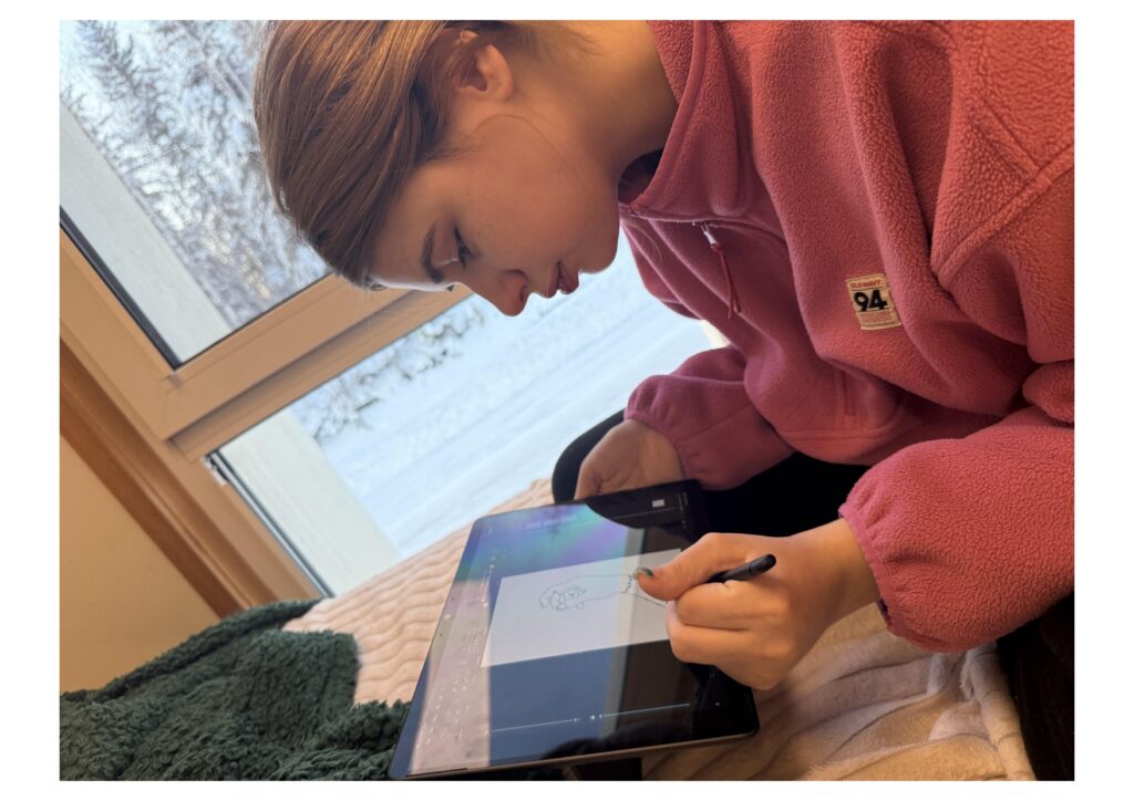
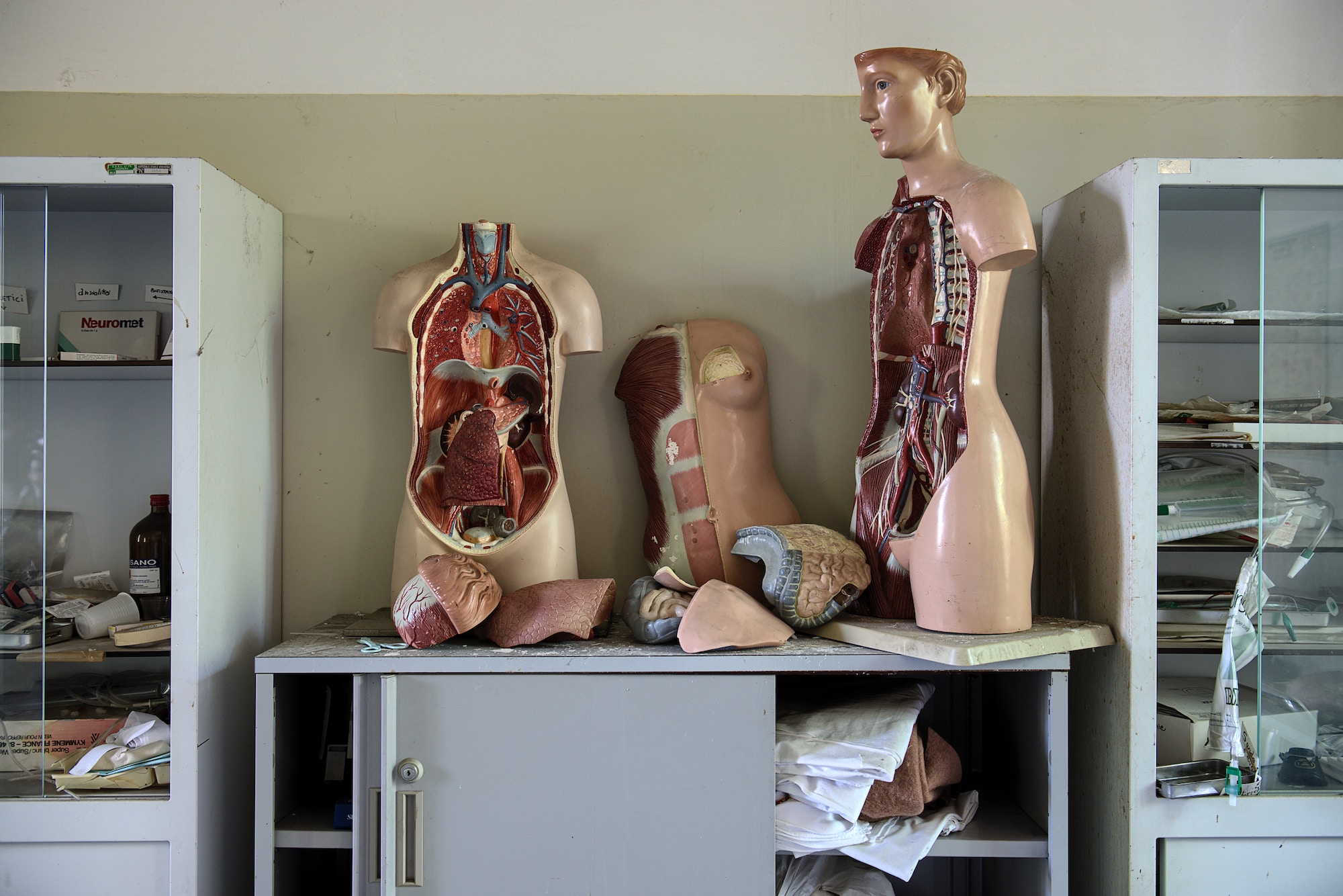
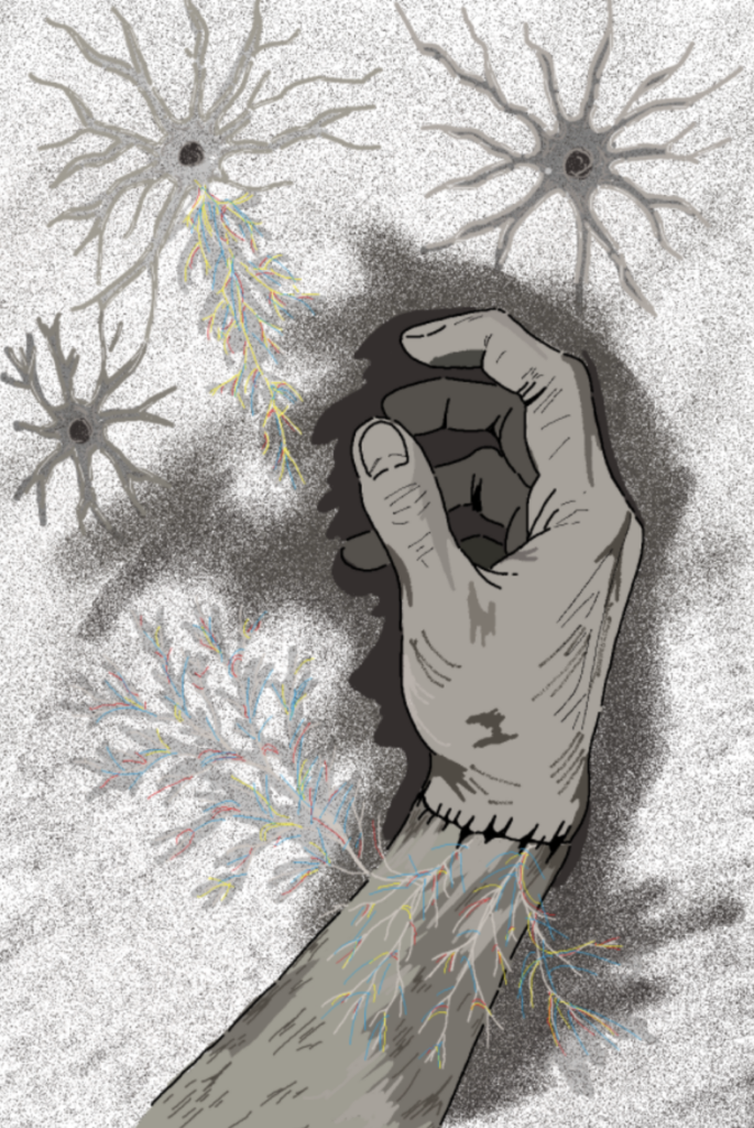
My artwork translates these physiological processes into symbolic visual language. Through anatomical drawing, I represent damaged nerve endings as dim and fractured pathways and regenerating axons as glowing threads of light. Schwann cells are depicted as protective structures guiding the nerves forward. Areas of synaptic reconnection appear as overlapping light patterns to show restored communication between neurons and muscles. I also used contrast between stillness and motion to reflect paralysis compared to recovery.
This project also fulfills the course objective of identifying key components and structures of the nervous system. Each drawing includes labeled anatomy such as axons, dendrites, Schwann cells, neuromuscular junctions, and muscle fibers. My goal is to showcase both scientific accuracy and emotional meaning by capturing the vulnerability and strength involved in rebuilding the body after trauma. Nerve regeneration is not only a clinical process but also a display of resilience at the cellular level.
Through this project, I have learned that recovery following limb reattachment is not a single event but a complex biological process involving injured neurons, supportive glial cells, chemical growth signals, and reactivated muscle tissue. The nervous system does not simply repair itself. It must rebuild and reconnect from microscopic foundations. By combining anatomy with my artwork, this project shows that science and humanity are truly inseparable in the process of healing.
References
Gordon, T. (2009). The role of neurotrophic factors in nerve regeneration. Neurosurgical Focus, 26(2), E3. https://pubmed.ncbi.nlm.nih.gov/19228105/
Liu, K., Tedeschi, A., Park, K. K., & He, Z. (2011). Neuronal intrinsic mechanisms of axon regeneration. Annual Review of Neuroscience, 34(1), 131–152.https://doi.org/10.1146/annurev-neuro-061010-113723
Sanes, J. R., & Lichtman, J. W. (1999). Development of the vertebrate neuromuscular junction. Annual Review of Neuroscience, 22, 389–442. https://doi.org/10.1146/annurev.neuro.22.1.389