 Posted by
Maggie Zaverl
Dr. Don Larson
Anatomy and Physiology II
31 Jul. 2025
Topic: Hypertensive Retinopathy
Medium: Drawing
Objective: “Know the path of blood through the heart and circulatory system”
During Unit 9, “Cardiovascular and Circulatory Systems”, the course covered the path that blood takes out of the heart and circles through the body. I would like to take this a step further and incorporate it into a topic that is important to me, eye health. My goal is to go to optometry school upon graduation from undergrad, and to help gain some experience, I’ve been working as an optometric technician for the past few years. In my time, I’ve seen many patients come through who have hypertension and we have to be vigilant in screening for hypertensive retinopathy. It’s important to keep an eye on blood pressure levels and keep up with medication, because eye health could be at risk, “Chronic-elevated blood pressure induces progressive alterations in the retinal vasculature, including arteriolar narrowing, arteriovenous nicking, and increased vascular tortuosity” (Souza 2025). When a patient has hypertension (high blood pressure), that high pressure in the body puts extra pressure on the vessels in the eye, which leaves them torturous, and can cause them to kink and break. After the vessel is broken, the tissue that that vessel was supplying blood to will die. After that, the tissue will atrophy or create new blood vessels, but these vessels will be leaky and weak. If they leak, the blood will seep into the retina (which is responsible for our perception of light, a.k.a. vision) in a process called neovascularization and could cause vision loss (Dr. Mavencamp). It’s important for hypertensive patients to have yearly dilated exams to assess for potential damage.
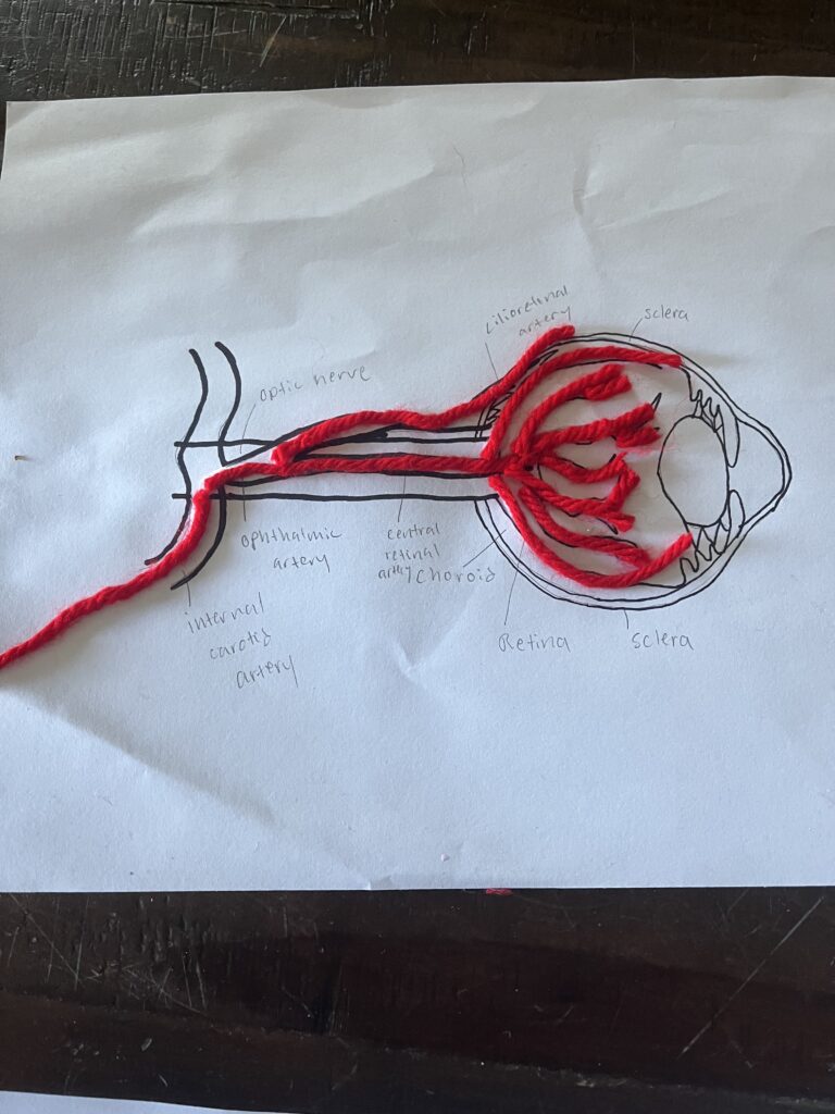
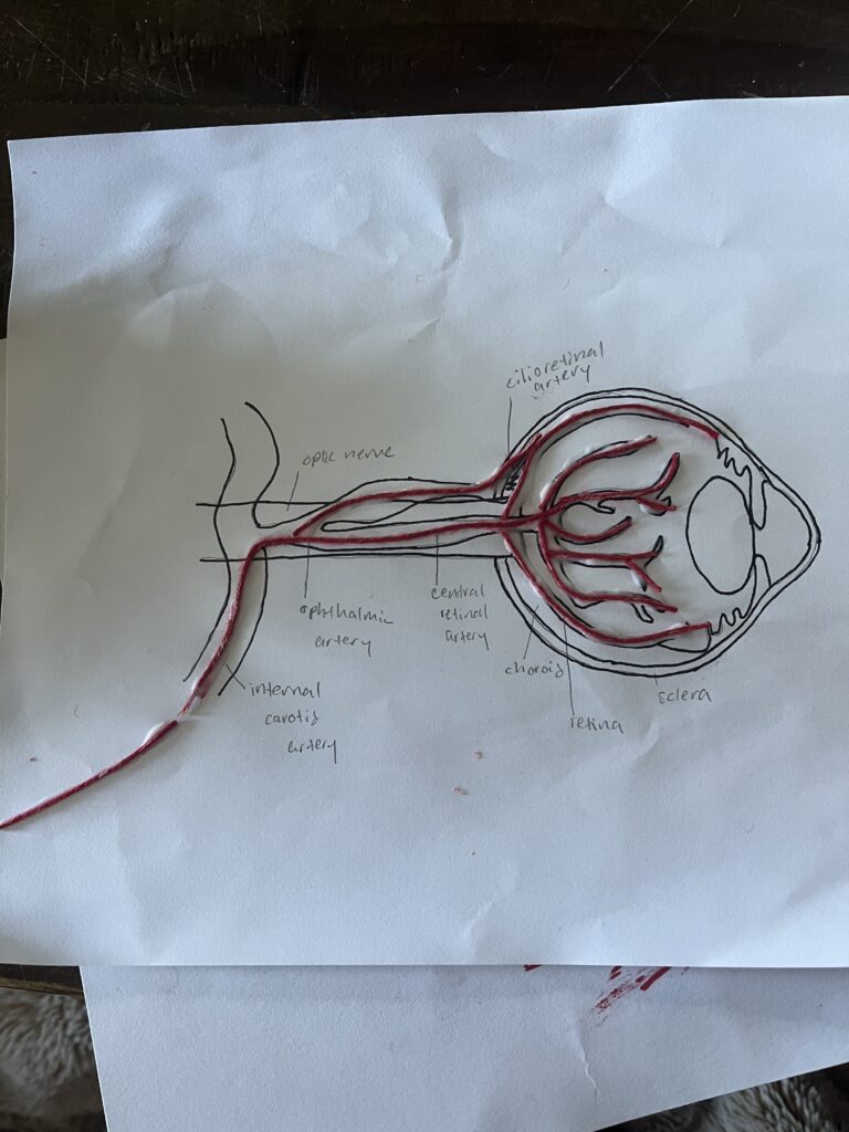
The path that blood takes from the heart to the eyes is important. It starts with the left ventricle and aorta: oxygenated blood is pumped from the left ventricle into the aorta. Then, the aorta sorts the blood using its many branches, and blood destined for the eyes flows through the branch going towards the head, mostly using the common carotid artery. The common carotid artery splits off into two branches on either side of the neck, the internal carotid artery and the external carotid artery. The internal carotid artery continues flowing into the skull to supply blood to the eye and brain. This is when the blood follows the internal carotid artery all the way into the ophthalmic artery. The ophthalmic artery is the main artery that supplies blood to the entire eye, so it’s very important. The ophthalmic artery has a few branches that deliver blood to the different parts of the eye. The central retinal artery follows the optic nerve to supply blood to the retina, which again, is responsible for observing light.
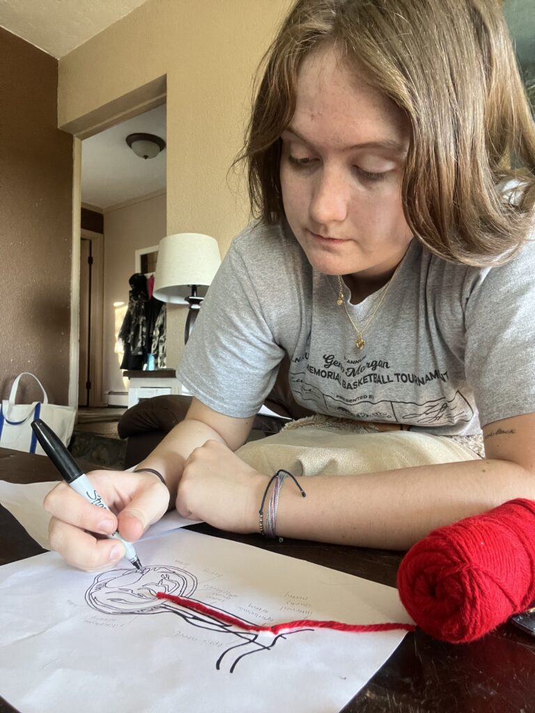
To demonstrate the path that blood takes to get to the eye, and the problems associated with the pressure that hypertension puts on the vessels of the eye, I drew two models of an eye: one with normal vessels (the string) and one with hypertensive vessels (the yarn). I used the thicker yarn to demonstrate hypertensive vessels because of the diameter changes vessels undergo when a patient has hypertensive retinopathy. The thicker vessels are what leads to the damage, “Hypertensive retinopathy (HR) refers to changes in the morphological diameter of the retinal vessels due to persistent high blood pressure” (Badawi et al. 2022). When the vessels get too big, and have too much pressure, they burst, and that’s when things go downhill. In my pictures, I also demonstrated the retina, the choroid (the layer between the sclera and and the retina which supplies nutrients and oxygen to the outer layer of the eye), the sclera (white of the eye) and the optic nerve (responsible for central vision and connection from eyes to brain). Patients who have hypertension might not think that their condition can cost them their vision, so it is crucial that they keep up with their treatment plans.
Sources
Badawi, S. A., Fraz, M. M., Shehzad, M., Mahmood, I., Javed, S., Mosalam, E., & Nileshwar, A. K. (2022). Detection and Grading of Hypertensive Retinopathy Using Vessels Tortuosity and Arteriovenous Ratio. Journal of Digital Imaging. https://doi.org/10.1007/s10278-021-00545-z
De Souza, E. C. (2025). Hypertensive Retinopathy. Diseases of the Retina and Vitreous, 1–7. https://doi.org/10.1007/978-3-031-29358-0_25-1
Dr. Michael Mavencamp, O.D. 2025.