 Posted by
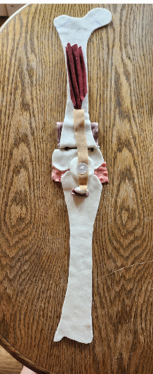
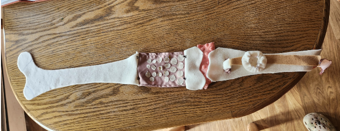
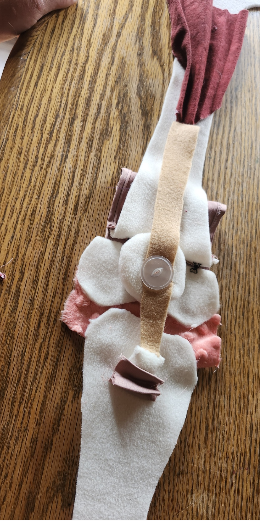
My project displays a femur, patella and tibia .The growth plate at the end of the femur can be open to see the different cell development zones. The growth plate on the tibial tuberosity can be observer thickening at the quadricep engages and pulls on the patellar ligament over the patella and to the tibial tuberosity which in an insertion point for that muscle. All materials are fabric and are hand sewn. All long bones there are epiphyseal plates, in this project I only focus on one on each bone. These are sites of growth in length on the long bones. On one side of the plate cartilage in formed on the other cartilage is ossified. There are four zones within the growth plate, the reserve zone, the proliferative zone, the zone of maturation and hypertrophy and the zone of calcified matrix. The reserve zone is closest to the epiphyseal end of the plate and is the home to small chondrocytes. These cells are not involved in growth but in securing the growth plate tissues to bone. Next is the proliferative zone, here larger chondrocytes reside in stacks, mitosis occurs here to make more chondrocytes to supply the next zone with more cells as they calcify and die. The older the cells are the closer to the diaphysis (shaft part of the bone) they are. Most of the growth in length happens in these two zones, the proliferation zone and the zone of maturation and hypertrophy. The zone of calcified matrix is similar to the first, in that its purpose is to secure the other end of the growth plate to the mature end of the diaphysis. However, it differs in that the cells are much larger here and the matrix around them has calcified (Waltzer). The growth hormone plays a predominant roll in the rate of growth in these plates and convert the plate into the epiphyseal line when that ensues no more longitudinal growth occurs. When injury or disease occurs in the areas it can have lasting effects on the development of the bone and the strength of the epiphyseal line in adulthood. Osteochondritis is a term the cover many disorders within the growth plates, that can stem from a disruption in the blood supply to the bone, necrosis and regrowth (Muchintala). In my project I compare a healthy growth plate to one with a type of osteochondritis, Osgood- Schlatter disease . In the healthy growth plate, the uniform nature of the different zones of cell can be observed. In the growth plate suffering from Osgood-Shaltters disease the cartilage is thickening because of the stress being put on it. The quadriceps the patellar ligaments insertion is on the tibial tuberosity with connects to the quadriceps. In and growing body there is a growth plate just under the tibial tuberosity, the strain of such big muscle group like the quadriceps pulling on the tuberosity can cause the chondrocytes in the proliferation zone and the maturation zone to lay down more cells in response to the stress. This can cause inflammation and pain, but generally fades with rest and ice and most people stop having recurrences when growth plate closes (Tokoph).
Muchintala, R., Coladonato, C., Perez, A., Kellish, A., Mumtaz, S., Sutton, W., Wilson, S., Cohen, S., Tjoumakaris, F. P., & Freedman, K. B. (2025). Return to Sport After Treatment of Stable Osteochondritis Dissecans Lesions of the Knee in Adolescents: A Systematic Review. American Journal of Sports Medicine, 53(7), 1761–1768. https://doi-org.uaf.idm.oclc.org/10.1177/03635465241272464
Tokoph, K. (2021, May 7). Apophyses of Long Bones, Osgood-Schlatter Syndrome, & Sever’s Disease [Video]. Youtube.com. Retrieved June 18, 2025, from https://www.youtube.com/watch?v=ff59RyIBBDI&t=473s
Walzer, S. M., Cetin, E., Grübl-Barabas, R., Sulzbacher, I., Rueger, B., Girsch, W., Toegel, S., Windhager, R., & Fischer, M. B. (2014). Vascularization of primary and secondary ossification centres in the human growth plate. BMC Developmental Biology, 14(1), 1–21. https://doi-org.uaf.idm.oclc.org/10.1186/s12861-014-0036-7