
Tooth Anatomy and Function
My STEAM project is on teeth, specifically their anatomy and function. The correlating topic in our course that relates to teeth and their function is the objective, “Know the structure of the knee joint and compare this joint type to other 5 joint types.” This may surprise you as most people consider teeth to be bones, but they are a type of joint!
The knee joint is the largest joint in our body, (Betts et al., 2022). Something we do not cover in depth is one of the smallest joints, which might surprise you: gomphosis. Gomphosis is a specialized fibrous joint that anchors the root of a tooth into its bony socket within the upper and lower jawbone in the skull, (Betts et al., 2022). Now that I have discussed my topic and how it relates to the objective, I want to discuss what makes up the anatomy of a tooth, the different types of teeth that are in human mouth and what each tooths specific function is.
The anatomy of a tooth has several different parts or layers. The first layer you can see is the crown of the tooth and the crown of the tooth is made of enamel. “Enamel consists of 96% mineral by weight primarily of a complex, highly organized structure of carbonate-substituted hydroxyapatite mineral arranged in interlocking prisms giving it its characteristic strength,” (Morris & Tadi, 2023). Enamel is harder than the bones in your body because it is made of calcium phosphate but unlike bones enamel does not regenerate. “STEM studies have shown that at the nanoscale, enamel comprises tightly bunched oblong crystals that are about 1,000 times smaller in width than a human hair. These tiny crystallites are made mostly of a calcium- and phosphate-based mineral called hydroxyapatite,” (Complexity of Human Tooth Enamel Revealed at Atomic Level In, 2020). According to Healthline magazine, based on the Mohs scale teeth are just as hard if not harder than steel. Your teeth are made to be incredibly powerful which is why they are so durable and perfect for mastication.
The next layer in a tooth is the dentin. Dentin makes up the core of the entire tooth that surrounds the pulp, which contains the neurovascular structures, (Morris & Tadi, 2023). The pulp is the life source of the tooth. The pulp contains all the blood vessels and nerves that allow your tooth to feel. Sometimes people get dental caries that destroy enamel and dentin and enter the pulp causing infection, (Huang, 2011).
The layer at the end of the tooth is called cementum. “The cementum is a layer of hard tissue that covers the root of the tooth. It is roughly as hard as bone but considerably softer than enamel. The connective tissues attach to the periodontal ligament, and through this bind the roots of the tooth to the gums and jaw (alveolar) bone,” (VC Dental, 2020). This binding is part of the “joint” of the tooth.
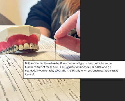
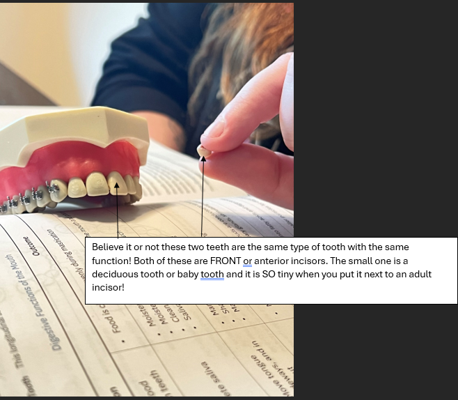
Each tooth in our dentition has a specific purpose and function. “Teeth provide a masticatory system that functions in the incising, tearing and grinding of food,” (Morris & Tadi, 2023). Different teeth can assist us in eating foods in different ways. As babies we have 20 primary or deciduous teeth. Babies have four molars, four incisors, and two canines. Incisors are meant to bite pieces of food, canines help tear food apart, and molar’s primary purpose is to grind the food down. Deciduous teeth do fall out and are eventually replaced by the adult dentition. Adults have twelve molars, eight bicuspids (premolars), four canines, and four incisors in a full dentition. Your front teeth which are referred to as central incisors don’t have roots that are as deep as other teeth and are usually singular in their roots. This means your front teeth aren’t as strong as other teeth because they only have one root as opposed to some molars who have two roots or more. Your anterior teeth are generally just for aesthetics. Your canines have the longest roots out of all your teeth and they’re usually wider and thicker making them suitable for ripping and tearing food. Molars are deep rooted with multiple roots and have a wider surface on the top of the tooth. Your molars are your biggest teeth and that is what makes them perfect for grinding food. Molars are also anatomically made for the stress and impact of your bite.
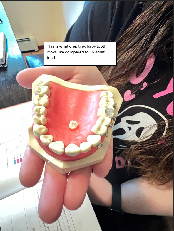
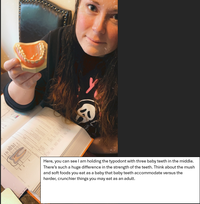
For my art piece I borrowed my wife’s typodont. A typodont is a set of teeth dental professionals use in school to learn and practice dental procedures. She has been a dental assistant for over 8 years. She also brought home some deciduous teeth for me to use after she sterilized them. I put the deciduous teeth inside the typodont of adult teeth to show the size and shape difference between baby teeth and adult teeth. I also wanted to show the different types of teeth so we took several photos of the typodont so you can see how each tooth looks.
References:
Morris, A. L., & Tadi, P. (2023, July 24). Anatomy, Head and Neck, Teeth. StatPearls – NCBI Bookshelf. https://www.ncbi.nlm.nih.gov/books/NBK557543/
VC Dental. (2020, January 13). Tooth Anatomy – Gosford, Experienced Dentists: VC Dental. https://www.vcdental.com.au/tooth-anatomy-education/#:~:text=The%20primary%20set%20of%20teeth,apart%20and%20molars%20grind%20food.
Betts, J. G., Young, K. A., Wise, J. A., Johnson, E., Poe, B., Kruse, D. H., Korol, O., Johnson, J. E., Womble, M., & DeSaix, P. (2022, April 20). https://openstax.org/books/anatomy-and-physiology-2e/pages/9-2-fibrous-joints. https://openstax.org/books/anatomy-and-physiology-2e/pages/10-4-nervous-system-control-of-muscle-tension
Kim, J., Lee, G., Chang, W. S., Ki, S. hyoung, & Park, J.-C. (2021). Comparison and Contrast of Bone and Dentin in Genetic Disorder, Morphology and Regeneration: A Review. Journal of Bone Metabolism, 28(1), 1–10. https://doi.org/10.11005/jbm.2021.28.1.1
Huang G. T. (2011). Dental pulp and dentin tissue engineering and regeneration: advancement and challenge. Frontiers in bioscience (Elite edition), 3(2), 788–800. https://doi.org/10.2741/e286
Complexity of human tooth enamel revealed at atomic level in. (2020, July 1). National Institutes of Health (NIH). https://www.nih.gov/news-events/news-releases/complexity-human-tooth-enamel-revealed-atomic-level-nih-funded-study
While reading Nikki’s post I learned about the anatomy and function of our teeth. They are a type of joint in our body. We identify the knee as the largest joint in our body but no one seems to identify in great detail the smallest joint in our bodies. That joint it Gomphosis. I learned that gomphosis is a specialized fibrous joint that anchors the root of the tooth into it’s bony socket within the upper and lower jawbone in the skull. We learn here that the anatomy of the tooth has many different layers. The first layer is the crown of the tooth which is made up of enamel. The second layer in the tooth is dentin which makes up the core of the whole tooth that surrounds the pulp. That contains neurovascular structures. The final layer, which is located at the end of the tooth is Cementum. This is the hard tissue that covers the root of the tooth. It is as hard as bone but it is much softer than enamel. There are connective tissues that attach to the periodontal ligament and this will bind the roots of the tooth to the gums and jaw. Also known as the alveolar bone.
I learned about the dentition in the each tooth. The different teeth in our mouth can aid us in eating food in different was. I learned that babies have 20 primary teeth. You molars, four incisors and two canines where as the adults have twelve molars, eight bicuspids, four canines and four incisors in a full dentition. The canines are the longest roots out of the teeth and the molars are the strongest as they are rooted deep with multiple roots and have a wider surface on top of the tooth. Anterior teeth are for the aesthetics.