
Just like any part of our body, teeth and their functions are important and play crucial roles. For my STEAM project, I decided to go further into the anatomy of teeth and oral cavity. And as I did my research, I found that teeth and bones are very much their own categories but they do relate to each other when it comes to the certain formation/ layers and their functions. For this art and research project, I’m going to be including the main objective: the functions of our bones and how they relate to our teeth and the oral structure.
A few main questions to think about during this relation is: What holds our teeth in place? And what protects our teeth?
Just like bones, teeth have major layers from inner to outer. But since there’s many of them, I decided to only talk about the important ones. I chose the ones that are crucial to the tooth’s structure and saw how their functions relate to our bones’. Teeth have eight main layers for protection and creation of the tooth including: the enamel, dentin, pulp, gums, cementum, root canals, periodontal ligament, and the alveolar bone.

For my art project, I started off with a blank canvas and painted it light pink.
Enamel is the outer layer we see in our mouth. It’s known as the strongest thing in our body, even stronger than bones, because of a mineral called hydroxyapatite. And just like bones, they are composed of other minerals like calcium and phosphorus. And even though bones have hydroxyapatite too, teeth have more of it which is what makes them as powerful as they are. But since the inner parts of our teeth are weak, we need enamel to provide defense against everyday use like bacteria and acids from the foods we eat. Bones also have a protective layer called the periosteum but this layer has other functions like supplying blood, growing, and repairing itself. Teeth cannot repair themselves.
Our tooth’s periodontal ligament is the layer after the cementum and basically has the same function. This area contains very soft connective tissue fibers. These fibers help secure the tooth/ teeth to the alveolar bone. They are also crucial to the integrity of our tooth’s socket due to helping it in the needed non-mobility. They aid in maintaining the cells that support and form our bone and cementum. The cells that are included are the fibroblasts, which help the revolution of the collagen fibers. This layer also contains the cementum cells called cementumblasts/ cementoclasts, which are just like our bones’ osteoblasts/ osteoclasts. The blasts help regenerate the structure, while the clasts break it down. And like I said before, I found that teeth can’t regenerate themselves but these cells are more crucial for the healing nature of our alveolar bone.
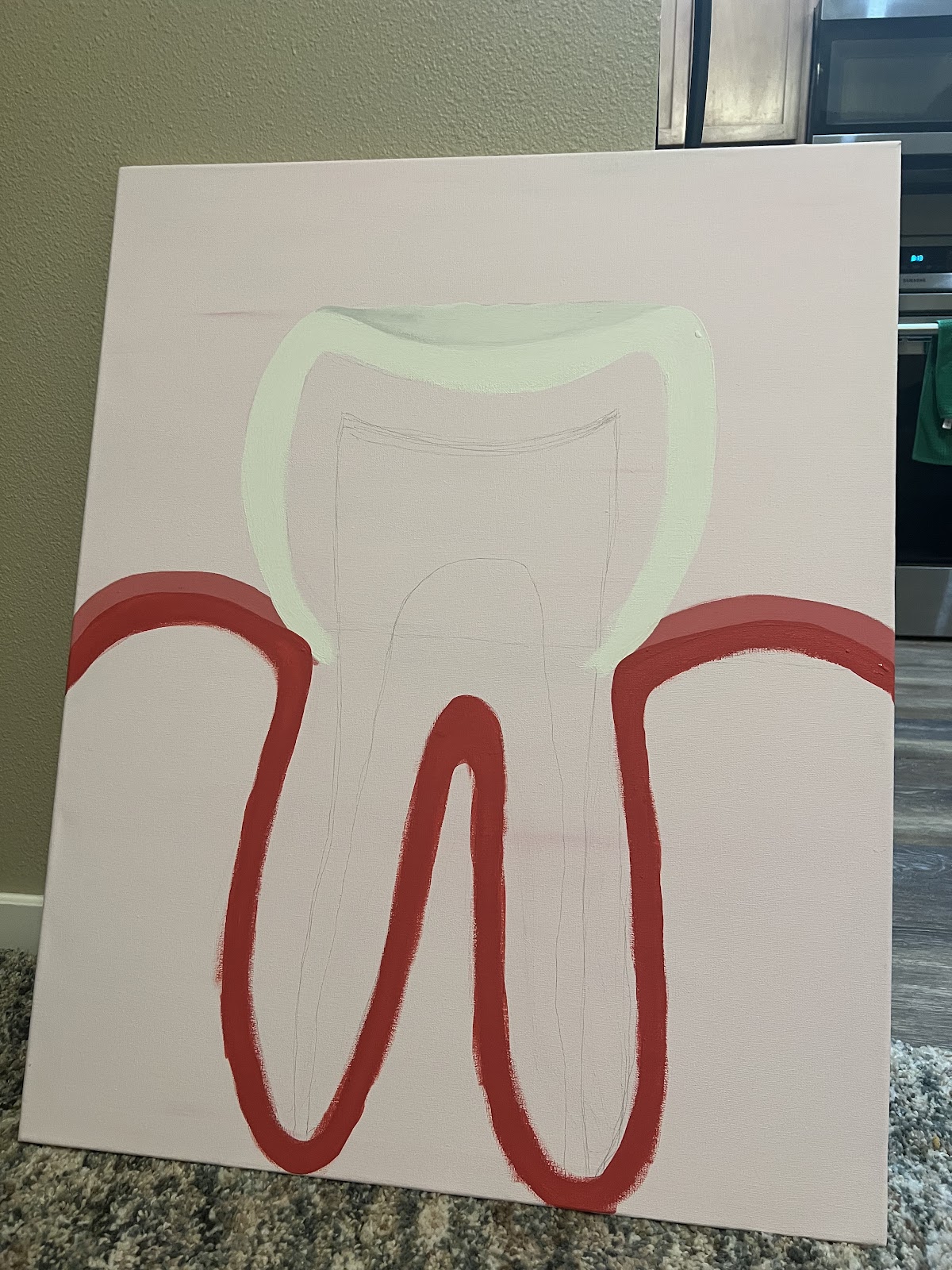
I then painted/ added the enamel, gum, and periodontal ligament layers.
Teeth have three main layers for protection: enamel, dentin, and cementum. But dentin is crucial for the tooth’s structure/ shape because this is what the enamel lays upon. It is also important for shielding the rest of the tooth because things can still break down our tough enamel. Dentin is used for the protection of our pulp and even distribution of pressure to prevent fractures. While the enamel does the same thing but is for just the protection of the outside of the tooth. And if there is any breakage, we need all the help we can get because the pulp is the weakest part. This layer is also very calcified like our bones, which is what makes them strong and good for the overall protection of our body and teeth.
The function of pulp provides things for our teeth like formation, senses, and nutrition. This layer includes the vessels that pump blood, nutrients, and odontoblast cells through our teeth to help form the surface we call dentin. The pulp also contains nerves that help relay information to the rest of our body. This is meant for times when we fracture our teeth or have cavities, so feel the pain. In our bones, we basically have the same set up. Bones have nerves that serve our body the same way, for times when we break our arm or any other part. In a typical bone there are blood vessels too: a nutrient artery, periosteal and epiphyseal vessels. These vessels serve the same purpose of pumping yellow/ red bone marrow, blood cells, nutrients/ waste, and osteoblast cells to help form not just one layer, but the overall structure of bone itself.
The cementum is the thin, hard, extra layer that covers right on top of the pulp and protects it. It lays right underneath the dentin layer and just like the dentin/ bone, this layer is super calcified. Cementum is also important for the anchoring of the tooth to the jawbone. This layer can either be cellular or acellular but it depends on which part of this area we are talking about. Acellular cementum has the specific fibers that only obtain the function of holding down the tooth and connecting it to the gums. Cellular cementum has thick collagen fibers just like our bones. The collagen in our teeth aids in the connection to the alveolar bone. And even though teeth can’t completely regenerate, I found that in the event of resorption, cementum can make minor repairs. For our bones, we need collagen to help create the overall structure and sturdiness. Collagen is also important for the regeneration of bones because unlike teeth, they can heal themselves fully.
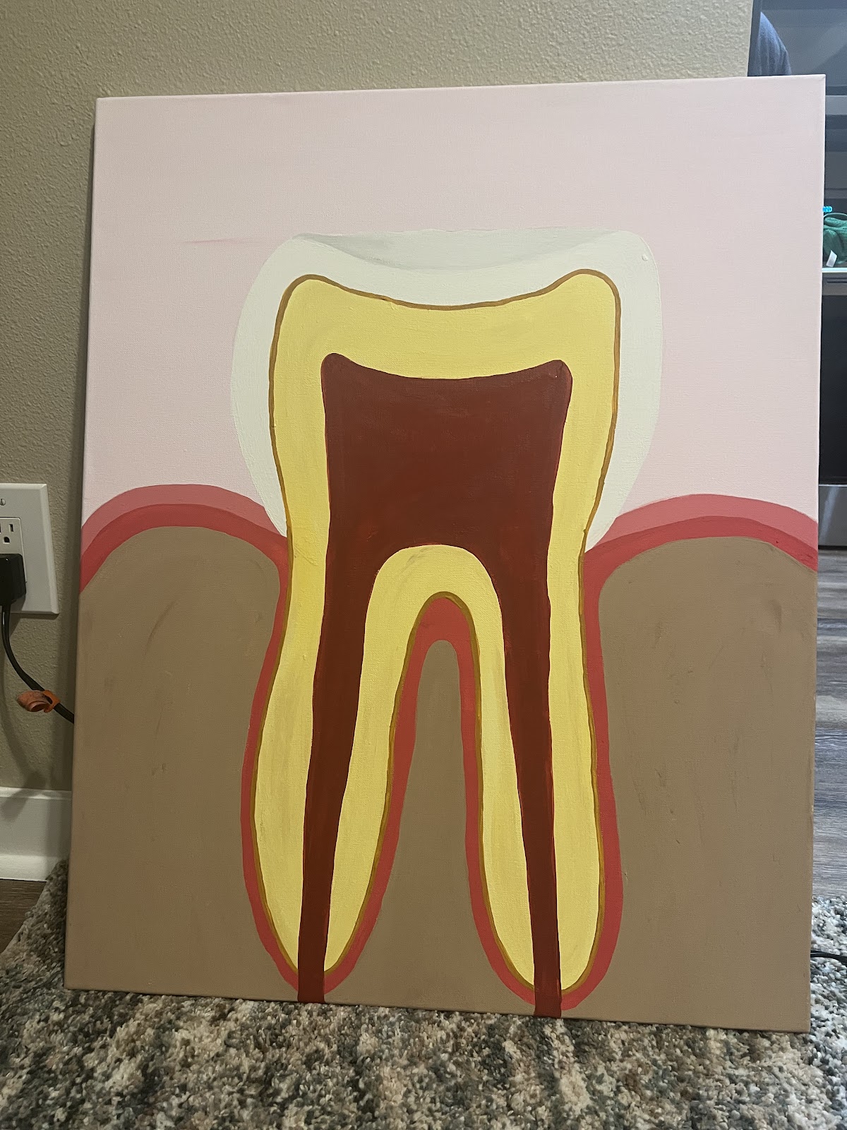
I then painted/ added the layers dentin, cementum, and the start of the pulp and two root- canals.
The root- canal is just the fancy word for pulp but just in a different part of the tooth. The pulp starts at the top of the tooth and runs throughout to the bottom, in the part we call the roots. Roots help stabilize the tooth and have the same nerves, blood vessels, and cementum as the rest of the structure. I found that the main function of root canals is just like the pulp. They aid in pumping nutrients and blood throughout the tooth, into the rest of the body. And just like bones, they help produce blood cells for our body to regulate our oxygen and nutrients. These blood cells from our teeth fight infections in the mouth. While the blood cells from bones fight infections for the rest of our body.
The alveolar bone, also known as the alveolar process, is the most important topic we have all been waiting for! This is the most relatable layer to our bones because it just simply is a part of our skeletal system. This thick bone has all the tooth sockets so it can provide the specific function of holding our teeth in the proper place. The periodontal ligament as well. This is needed so it can obtain certain pressures/ forces while doing actions like chewing. And since the alveolar process is just regular bone, it has basically the same functions of bone. The alveolar supports our teeth, protects the nerves/ vessels that circulate through the teeth, regenerates through osteoblasts, deteriorates through osteoclasts, and has calcium mineral storage. The only difference is that other major bones support our whole body, while the process just deals with the oral cavity.
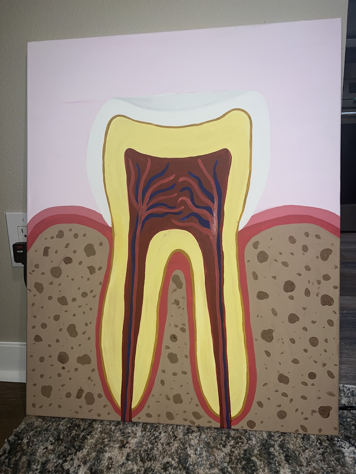
I then added brown spots to enhance the alveolar bone or process. I also added red and blue lines running through the pulp/ root- canals to show the nerves and blood vessels.
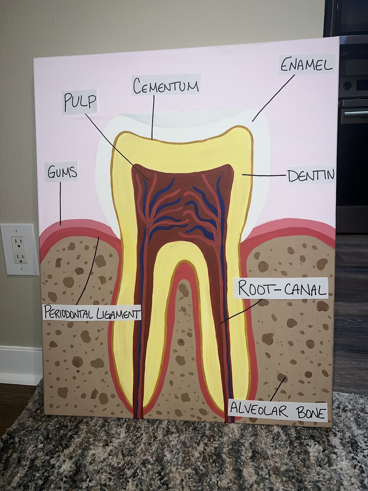
Then I finished it off by adding labels to each of the layers!
When I first started doing my research, I thought maybe my topic/ art was going to be hard to relate to my objective. This is because I found that teeth are teeth and not really classified as bones. Reasoning why is mostly because of their composition and where they are located in the body. Bones are living tissues that are strong, can regenerate themselves, and are located throughout the body to make our skeletal system. While teeth are only defined as non living, they can’t remake themselves, and can only be found in our mouth. But while having these major differences, these two can be one of the same by providing protection and growth for our body.
Sources
Woelfel’s Dental Anatomy. Rickne C. Scheid. Call No. QM311. W64 2012.
Development, Function, and Evolution of Teeth. Mark Franklyn Teaford, Moya Meredith Smith, and Mark W. J. Ferguson. Call No. QP88.6. Cambridge University Press, 2000.
Clinical Anatomy, “Anatomy in Dentistry- From the Beginnings to Contemporary Reality.” September 2022. Vol. 35 Issue 6, Pages 711- 722.
Journal of Morphological Sciences, “Tooth Anatomy in Dental Education: A Better Way to Replicate Dental Morphology.” 2023. Vol. 40, Pages 215-219.
Anatomical Sciences Education, “Three- Dimensional Tooth Models with Pulp Cavity Enhance Dental Anatomy Education.” May- June 2022. Vol. 15 Issue 3, Pages 566-575.
Unit 4 Lecture Notes: Skeletal System. Don Larson. Publication Date, Unknown. Pages 1- 105.
Teeth: Anatomy, Types, Function & Care (clevelandclinic.org) Teeth: Anatomy, Types, Function, and Care. Cleveland Clinic. 2023.
Cementum – an overview | ScienceDirect Topics Principles of Regenerative Medicine, “Cementum – An Overview.” ScienceDirect. 2008. Periodontal Ligament and Alveolar Bone in Health and Adaptation: Tooth Movement – PMC (nih.gov) Front Oral Biology, “Periodontal Ligament and Alveolar Bone in Health and Adaptation: Tooth Movement. NIH: National Library of Medicine. 2016. Vol. 18. Pages 1-8.
Halie you did such an amazing job. Your descriptions are very clear and easy to understand. The artwork to go with is nicely laid out, so you can read, then look at the paintings and understand exactly what is going on. It directly relates to this course and explains even more in depth than we’ve gone over in relations to class currently. Your artwork is an accurate depiction and you can clearly tell you put time into the step by step process of your assignment.