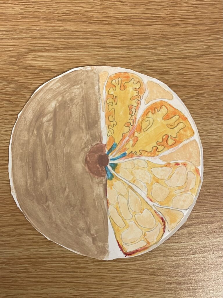
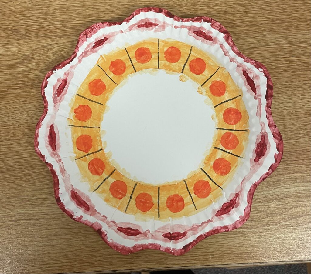
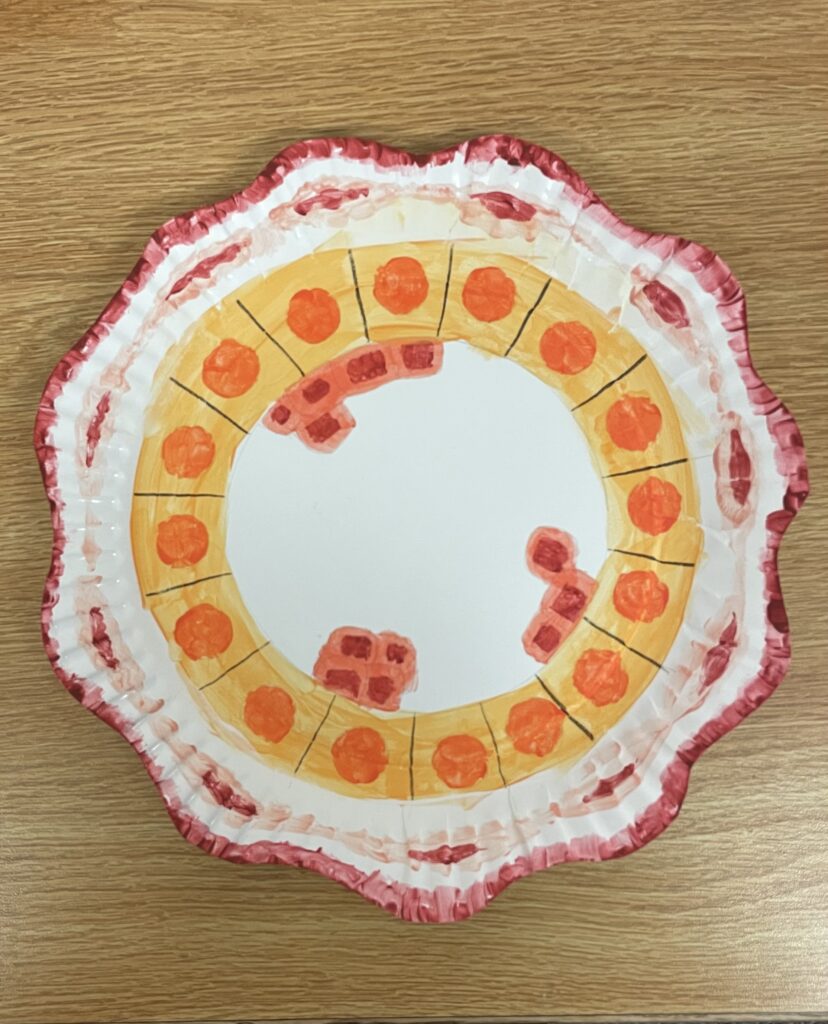
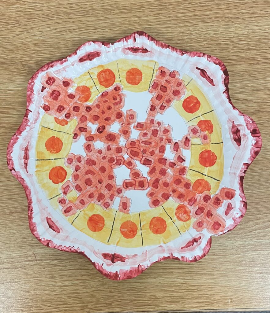
This is the breast and what is inside of it. The lower right portion of the photo shows the lobes and the upper right portion is what the inside of the lobe looks like. The blue parts of the illustration are the ducts.This is what a normal, healthy duct looks like. It is a tube-like part of the breast and this is where the lactation comes through before going out the nipple. This is the ductal hyperplasia and it also called the duct epithelial hyperplasia. The ductal hyperplasia is an growth of cells that line the small tubes inside of the breast. This is the atypical hyperplasia or the ADH and it affects the cells of the milk ducts in the breasts. This is not considered cancer, but it increases the risk of having breast cancer. DCIS or ductal carcinoma in situ is a cancer that affects the cells in the milk duct. The cells lining the milk duct become cancerous, but stay in the the situ. This is the early stages of breast cancer. This is the invasive ductal carcinoma and it grows in the ducts and invades the fatty or fibrous tissues of the breast outside of the milk ducts. The invasive ductal carcinoma is the most common type of breast cancer and can spread to other organs of the body.
One Comment
Autumn’s project was on breast cancer development. She stated that her objective was breast cancer development. She outlined this objective using painted plates as her media. These plates depict the breast in its entirety, a healthy duct, a duct with hyperplasia, a duct with atypical hyperplasia, a duct with carcinoma in situ, and a duct with invasive carcinoma. The first plate shows a breast in its entirety with the ducts painted in blue. The painting of the anatomy of the outside and the inside of the breast allowed the viewer to locate the area of the breast that would be discussed throughout this project. The second plate shows a healthy duct. This plate outlined the golden standard for a duct and aided in the viewer’s understanding of what a healthy growth-free duct looks like. The third plate shows a duct with hyperplasia. This plate showed the first sign of any unusual growth while still outlining that this type of epithelial growth does not affect much in the duct. The fourth plate showing the duct with atypical hyperplasia shows a growth that may affect the milk duct but is still not considered cancerous yet. The fifth plate shows a duct with carcinoma in situ or early breast cancer. This plate shows a growth that stretches across the milk duct. At this point, the growth of the cells occurring in the duct is vast enough to be considered cancerous. The sixth plate shows a duct with invasive carcinoma or advanced breast cancer. This plate shows extensive growth that expands beyond the milk duct, has entered other tissues and has possibly metastasized. These plates show the progression of a healthy milk duct to an advanced cancerous duct by displaying growths that occur within ducts at varying levels of health.


Autumn’s project was on breast cancer development. She stated that her objective was breast cancer development. She outlined this objective using painted plates as her media. These plates depict the breast in its entirety, a healthy duct, a duct with hyperplasia, a duct with atypical hyperplasia, a duct with carcinoma in situ, and a duct with invasive carcinoma. The first plate shows a breast in its entirety with the ducts painted in blue. The painting of the anatomy of the outside and the inside of the breast allowed the viewer to locate the area of the breast that would be discussed throughout this project. The second plate shows a healthy duct. This plate outlined the golden standard for a duct and aided in the viewer’s understanding of what a healthy growth-free duct looks like. The third plate shows a duct with hyperplasia. This plate showed the first sign of any unusual growth while still outlining that this type of epithelial growth does not affect much in the duct. The fourth plate showing the duct with atypical hyperplasia shows a growth that may affect the milk duct but is still not considered cancerous yet. The fifth plate shows a duct with carcinoma in situ or early breast cancer. This plate shows a growth that stretches across the milk duct. At this point, the growth of the cells occurring in the duct is vast enough to be considered cancerous. The sixth plate shows a duct with invasive carcinoma or advanced breast cancer. This plate shows extensive growth that expands beyond the milk duct, has entered other tissues and has possibly metastasized. These plates show the progression of a healthy milk duct to an advanced cancerous duct by displaying growths that occur within ducts at varying levels of health.