
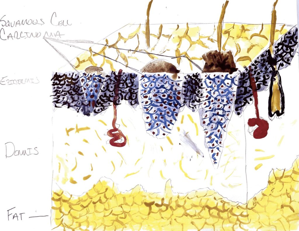
For my STEAM project I will be discussing squamous cell carcinoma of the skin. The objective form our class I will be expanding on is from our section on tissues, state location and various tissues of the body. Squamous cell carcinoma of the skin affects the epidermis of our skin where stratified squamous cells are located. The bottom layer of the epidermis is cuboidal where it gives way to stratified squamous cells. These cells become ketatinized and will eventually slough off, allowing for healthier tissues to take its place.
Excessive sunlight exposure is the main cause for squamous cell carcinoma. UV light emitted from the sun, more specifically UVA, UVB, and UVC are responsible for genetic mutations that result in the formation of cancerous cells. Our DNA is very capable of absorbing UV light and redistributing it as energy within the cell. With excessive UV exposures, a couple of things can happen that could lead to cancerous cells. As we learned in our section on chemistry, a Nucleic Acid has four bases: Adenine, Thymine, Guanine, and Cytosine. A pericyclic reaction is when the bases of Thymine and Cytosine are fused together causing the DNA repairing process to be inhibited, possibly leading to cancer. Another way that cancer can form is when our melanin, which also absorbs UV light and transfers it as energy to the cell, can redistribute to a part in the cell that cannot handle this energy. An amino acid or oxygen molecule are examples of structures in a cell that cannot retain this energy properly, which in turn becomes an excited molecule and could damage DNA in the cell.
Once a group of cells can no longer repair their DNA a process of aging can occur. The aging process makes the surface of the skin appear darker. This are of the skin is know more susceptible to further UV damage, which is why we see squamous cell carcinoma start in the top layer of the epidermis. It can migrate deeper in to the dermis and even layers of fat and bone if left untreated. As we learned in our tissues section and integumentary section, stratified squamous is the tissue that makes up all of our exterior skin leaving it the most susceptible to damage from UV light.
While we learned about tissues, my dad was diagnosed with squamous cell carcinoma of the skin, which was luckily found early by a dermatologist. Although it was not a fun call to get, it was helpful to learn about this subject in depth as my dad was going through this. A Mohs procedure was recommended to him because it is highly successful at eliminating squamous cell carcinoma of the skin, typically found on the head. Mohs is where the affected area is removed by a surgeon. The surgeon, going off of sight will remove enough tissues both circumference and depth of the affected area. The tissue is then sliced into extremely thin layers, starting from the top to the bottom of the tissue sample. It is through this process that the surgeon is able to see how far down into the epidermis the cancer spread by looking at each section of the tissue sample through a microscope. If the surgeon seen cancer in the bottom layer they will remove more tissue until no more cancer is found. I am happy to report that this procedure took a couple hours and resulted in my dad now being cancer free. For my art piece I decided to paint with water color, a section of the skin layers and what squamous cell carcinoma looks like compared to healthy stratified squamous cells.
References:
Schuch, A. P., Moreno, N. C., Schuch, N.J., Menck, C.F.M., & Garcia, C.C.M. (2017, January 18) Sunlight damage to cellular DNA: Focus on oxidatively generated lesions. Free Radical Biology and Medicie. Retrieved November 22, 2021, from https://www.sciencedirect.com/science/article/pii/S0891584917300382.
Nature Publishing group. (n.d.). nature news. Retrieved November 22, 2021, from https://www.nature.com/scitable/blog/scibyts/how_ultraviolet_light_reacts_in/.
Christian Murray, D.S. (n.d.). patient indications for Mohs micrographic surgery: A systematic review – christian murray, Duvaraga Sivajohanathan, Timothy P. hanna, Scott Bradshaw, Nowell Solish, Benvon Moran, Robert Hekkenberg, Alice C. Wei, Teresa Petrella, 2019. SAGE Journals. Retrieved November 22, 2021, from https://journals.sagepub.com/doi/full/10.1177/1203475418786208.
This project tackles squamous cell carcinoma of the skin. It uses a watercolor painting to help us visualize the carcinoma. In it we can see that it affects the epidermis, where the stratified squamous cells are located. We can see that the deeper layers become cuboidal. In the watercolor, we can see that the carcimona is darker then the surrounding skin, which we learn is due to damage from UV rays, either UVA, UVB, or UVC rays. These damage the genetic code of the cell, leading it to turn cancerous. As it darkens it becomes even more susceptible to further damage. As it grows it deepens and left untreated can even reach layers of fat or bone. We also learn that there is a procedure, in this case a Mohs procedure, to remove the carcinoma where a surgeon carefully cuts around the tumor. A microscope is used to examine the bottom layers, and the surgeon continues to cut until no further cancerous cells are found.