
My STEAM Project focused on the largest organ of the human body, the skin. Personally, I am particularly curious as to how it remains strong and resilient despite the many breaches humans subject it to. My project aims to describe tattoos and its effect on skin tissues and other related parts. Specifically, my project’s aim is to describe tissues in the integumentary system and their functions. Moreover, I hope that through my project, I am able to provide a simple and straightforward interpretation of the information I was able to gather.
Depending on which reference you are using, tattooing has a long history that encompasses its early history and the instruments in its application. For the longest time, it has been attributed to the Samoans however this was debunked by the discovery of the Iceman, the most famous archaeological evidence of tattoos, 59 tattoos in all. But many researchers continue to believe that perhaps many more remain from various areas around the world. Its symbolisms have likewise changed over the years from marks of high status to self-expression. They were originally applied using sharp instruments from the surroundings like animal bones and the ink made from ground charcoal.
Independent research startup firm Statista Research found out that tattoos, along with piercings and plastic surgery are the most common modifications in the United States. Their current ‘statistics’ did also mention that about 29% of Americans (population was only 1,021 though). According to the American Academy of Dermatology, 24 percent of people from ages 18 to 50 have a tattoo (Betts, Young, Wise, Johnson, Poe, Kruse, Korol, Johnson, Womble, & DeSaix, 2020).
The process of how the tattoo penetrates our skin can be simply described vis-a-vis the skin’s two main layers: epidermis, which forms a protective barrier against infections and made up of closely packed epithelial cells and the dermis, which contains the hair follicles, sweat glands and blood vessels and made up of dense, irregular connective tissue. As with any foreign object, the body reacts to it and in this case, sends a horde of white blood cells to remove them. Tattoo needles have at least three sharp points for fine lines, and many more for shading. Through capillary action, the ink is held in place between these needles. Through a combination of adhesive forces and surface tension, the ink pushes through the skin until it reaches the dermis. The affected areas will then swell and turn red and eventually begin to scab over and flake. However, the ink particles are much larger than white blood cells, and while some will be removed, the majority will remain on the skin.
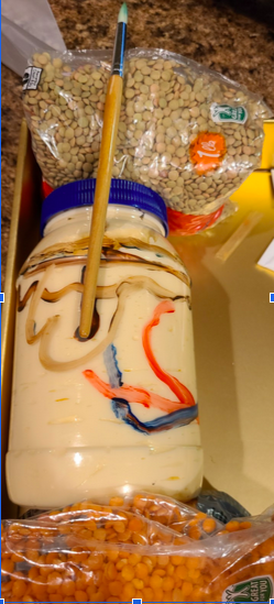
My project highlights the image representation (Schreiver, Hesse, Seim, Castillo-Michel, Villanova, Laux, Dreiack, Penning, Tucoulou, Cotte, and Luch, 2017) of translocation of tattoo particles from skin to lymph nodes. This simple image highlights the fate and effects of tattoo pigments in human skin. To differentiate the layers, I decided to use various types of ‘layers’ in my own apartment. The image depicts and highlights the effect of tattooing and its extent to the skin and skin tissues. The different layers and the respective materials I used are as follows: the epidermis was represented by the lentils, shaded with the particles of pigmentation. The red lentils on the other end are the hypodermis, with the dermis layer between them represented by the mayonnaise, which doubled as a canvas for further illustration. The red and blue lines represent the vascular connections within the dermis and to the root of the hair, which is represented by the brush handle and extends on the opposite end above the epidermis, with the brush end representing the frayed and damaged hair tip. Figure 1 below is the inspiration for the illustration.

Figure 1. Adapted from Schreiver, I., Hesse, B., Seim, C., Castillo-Michel, H., Villanova, J., Laux, P., Dreiack, N., Penning, R., Tucoulou, R.,Cotte, M., and Luch, A. (2017). Synchrotron-based v-XRF mapping and μ-FTIR microscopy enable to look into the fate and effects of tattoo pigments in human skin. Scientific Reports, 7(11395). https://doi.org/10.1038/s41598-017-11721-z
While very technical and beyond my understanding, I believe that the study which used synchrotron μ-FTIR analysis on skin and lymphatic tissues of human corpses from the Institute of Forensic Medicine at the Ludwig-Maximilians University of Munich. Further advanced methods like advanced mass spectrometry revealed a “simultaneous transport of organic pigments, heavy metals and titanium dioxide from skin to regional lymph nodes.” The study further highlighted organic pigments reaching the lymph nodes more. Ultimately, their study was able to emphasize through deep-penetration x-ray “ultrastructural changes of the tissue adjacent to tattoo particles through altered amide I α-helix to β-sheet protein ratios and elevated lipid contents (Schreiver, Hesse, Seim, Castillo-Michel, Villanova, Laux, Dreiack, Penning, Tucoulou, Cotte, and Luch, 2017). In simple terms, it further stresses the placement of toxic elements that contribute to negative effects of tattoos. This is similar to an earlier study by Grant, Twigg, Baker, & Tobin (2015) which examined tattoo ink nanoparticles in skin tissue and fibroblasts using atomic force microscopy (AFM). The results revealed that the nanoparticles of the tattoo ink are still detectably present in both skin tissues and fibroblasts. In addition, the fibroblasts were said to not likely be permanently damaged by the tattoo. On the other hand, the skin tissue analysis revealed a redistribution of the pigments deeper into the dermis and some even entering dermal blood vessels before transportation to lymph nodes (Grant, Twigg, Baker, & Tobin, 2015).
At present, tattoos are ubiquitous in modern society; however, they do not come without risk of medical complications (Winn, RIvard, and Green, 2016). Typical (and expected) complications include allergic reactions (to the ink); skin infections (from the instruments); blood-borne diseases, such as tetanus, hepatitis C, and hepatitis D; and the growth of scar tissue. A person with tattoos should be cautious when having a magnetic resonance imaging (MRI) scan because an MRI machine uses powerful magnets to create images of the soft tissues of the body, which could react with the metals contained in the tattoo dyes (Betts, Young, Wise, Johnson, Poe, Kruse, Korol, Johnson, Womble, & DeSaix, 2020). Considered as a form of skin trauma, it should heal via local treatment and with no infection and no local complication (Serup, 2017). Its removal, like its application, still poses an effect to the skin. Pulsed light and laser are still typically used for tattoo removal.
References:
1) Schreiver, I., Hesse, B., Seim, C., Castillo-Michel, H., Villanova, J., Laux, P., Dreiack, N., Penning, R., Tucoulou, R.,Cotte, M., and Luch, A. (2017). Synchrotron-based v-XRF mapping and μ-FTIR microscopy enable to look into the fate and effects of tattoo pigments in human skin. Scientific Reports, 7(11395). https://doi.org/10.1038/s41598-017-11721-z
2) Basel, K. (2017). Diagnosis and therapy of tattoo complications. With atlas of iIllustrative cases. (Serup, J. & Bäumler, W, Eds), Curr Probl Dermatol. vol 52, pp 74-81. https://doi.org/10.1159/000450804
3) Diogo Oliveira. (2008, June 13). Strange Science – Tattoos [Video]. YouTube. https://youtu.be/Id2i4Z1rwVU
4) Gordon Betts, J., Young, K., Wise, J., Johnson, E., Poe, B., Kruse, D., Korol, O., Johnson, J., Womble, M., DeSaix, P. (2020). Anatomy and Physiology. Houston, Texas: OpenStaxs. Retrieved from https://openstax.org/books/anatomy-and-physiology
5) Grant, C. A., Twigg, P. C., Baker, R., & Tobin, D. J. (2015). Tattoo ink nanoparticles in skin tissue and fibroblasts. Beilstein journal of nanotechnology, 6, 1183–1191. https://doi.org/10.3762/bjnano.6.120
Joshua’s project was centered around the integumentary system and the effects that tattooing has on it. As someone with a handful of tattoos, I found this interesting right off the bat. Tattoos are a major form of self-expression, dating all the way back to the Iceman, where they used tools like sharp bones and ground charcoal. Nowadays, tattoos are considered to be one of the most common forms of body modifications. Tattoo needles breach the skin until they reach the dermis, where the majority of ink will stay, and some will be removed by white blood cells. In this project, house-hold items were used to imitate the layers of the skin, and show how the needles disperse ink into the dermis. Included with this were also some possible side effects from tattooing like allergic reactions, skin infections, and hepatitis C & D. Tattoos are considered open wounds and skin trauma, so they need to be treated delicately and they will heal properly.