This project is a representation of the bone structure and development. The fetus represents an infant in the womb, whose bones are primarily composed of cartilage instead of the spongey bone matrix. Once the fetus is born, the bone turns into a true bone structure with the help from the osteoclasts and osteoblasts (blue and green blobs). The bone structure is like a brick wall, where layers are always being broken down and built back up. The bone is supplied by blood vessels and surrounded by nerves that run in and out of cavities within the bone. 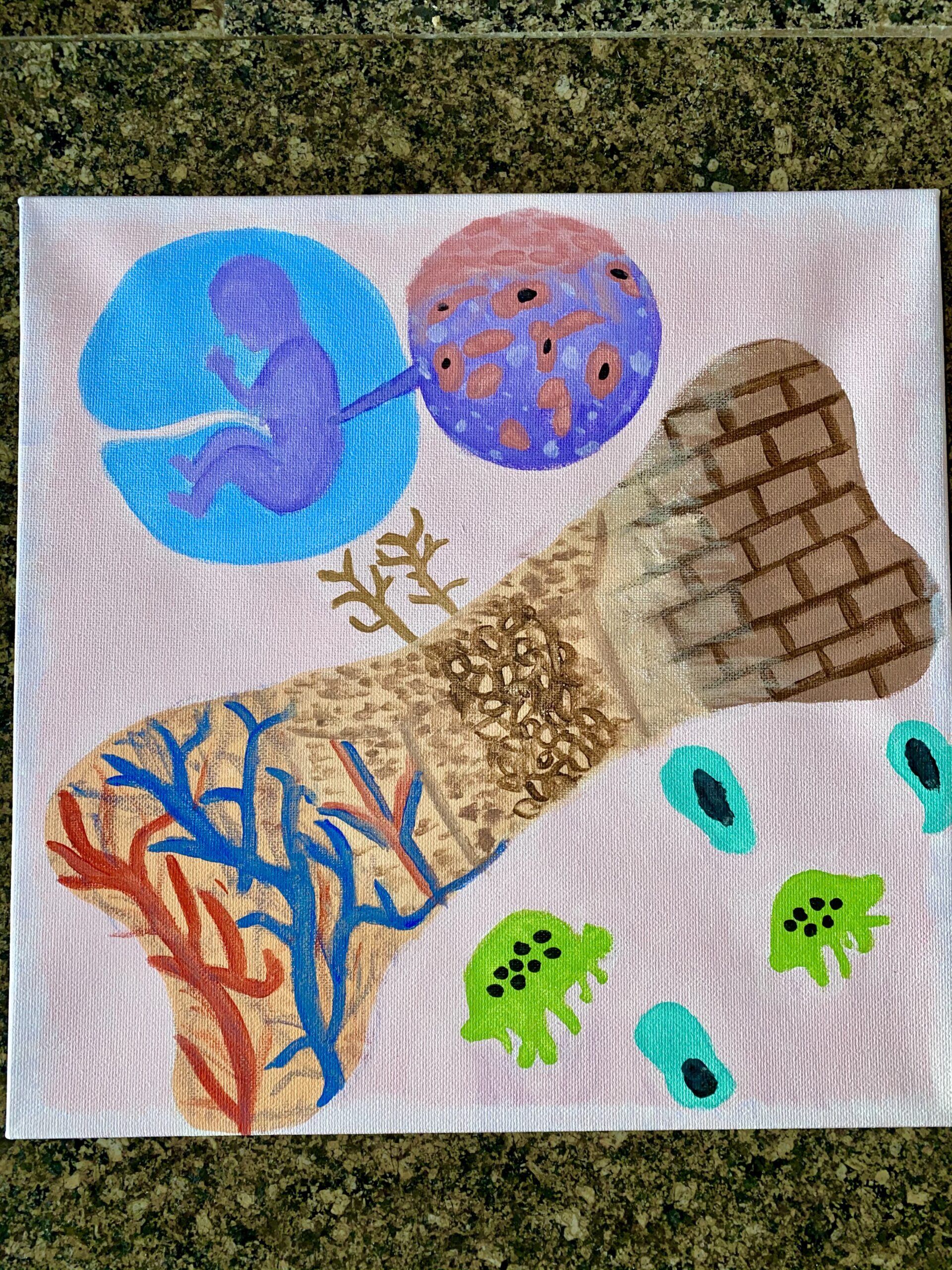

The process of bone development begins with the fetus in the womb. A “blueprint” of sorts is lied out and formation begins. First hyaline cartilage—a semi-solid matrix—is created to begin forming the fetal skeleton. The hyaline matrix is made up of chondroblasts, chondroitin sulfate, hyaluronic acid, collagen, and water. As the baby continues to develop, their skeleton will turn from hyaline cartilage into true bone. Some of their skeleton (i.e., the top of their skull) remain as soft cartilage until further development occurs.
Some functions of bones are; support, protection, movement, mineral growth, and storage, blood cell creation, fat storage, and hormone regulation. In order to stay in good condition, bones must go through a remodeling process. Adjustments must be made to them throughout our lives. The process of bone remodeling begins with resorption by osteoclasts. Then osteoblasts come in in order to lay down the foundation for new bone growth. This process is heavily influenced by hormone regulators such as; parathyroid hormone, calcitrol, and growth hormones like glucocorticoids, sex hormones, and thyroid hormones. Abnormalities can happen with bone remodeling. These abnormalities can lead to diseases such as cancer.