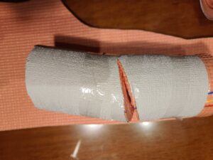
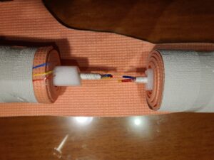
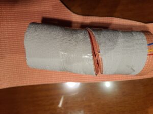
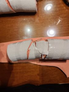
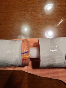
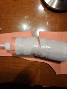
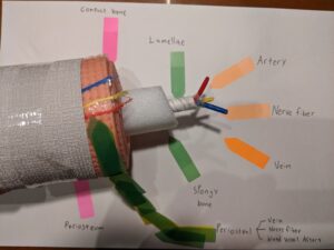
In Pete’s project, he included ten images to represent his sculpture. It was of the different forms of a stress fracture in the bone. Pete constructed a 3D model of a bone with the middle of the sculpture representing the lamellae layer, spongy bone layer, blood vessels, arteries, and nerves. The outside was the compact bone, periosteum, and periosteal layers. The first image was that of an open fracture, this is when the bone protrudes from the body and is exposed. The second image shown is a closed fracture, similar to the open fracture but the bone remains in the skin. The different classifications of a fracture are open fracture, complete fracture, closed fracture, and incomplete fracture. Each open fracture and closed fracture can be either incomplete or complet4eImage three shows that of an incomplete fracture because the bone is still connected. Image four is that of a complete fracture because the bone has been completely severed and separated. Image 6 is closed and incomplete because no skin was opened and the bone did not separate. Image seven was open and incomplete because the skin was opened, exposing bone but the bone did not separate completely. Image eight was a closed fracture that was completely separated. The other two types of classification that Pete showed were displaced fracture and nondisplaced fracture. The displaced fracture means that the alignment is not in its usual position while the nondisplaced means the alignment is still correct. Image nine and ten represented these classifications. Nine being nondisplaced and image ten was a displaced fracture. His project was easy to follow and created a great image for each form of bone fracture.


In Pete’s project, he included ten images to represent his sculpture. It was of the different forms of a stress fracture in the bone. Pete constructed a 3D model of a bone with the middle of the sculpture representing the lamellae layer, spongy bone layer, blood vessels, arteries, and nerves. The outside was the compact bone, periosteum, and periosteal layers. The first image was that of an open fracture, this is when the bone protrudes from the body and is exposed. The second image shown is a closed fracture, similar to the open fracture but the bone remains in the skin. The different classifications of a fracture are open fracture, complete fracture, closed fracture, and incomplete fracture. Each open fracture and closed fracture can be either incomplete or complet4eImage three shows that of an incomplete fracture because the bone is still connected. Image four is that of a complete fracture because the bone has been completely severed and separated. Image 6 is closed and incomplete because no skin was opened and the bone did not separate. Image seven was open and incomplete because the skin was opened, exposing bone but the bone did not separate completely. Image eight was a closed fracture that was completely separated. The other two types of classification that Pete showed were displaced fracture and nondisplaced fracture. The displaced fracture means that the alignment is not in its usual position while the nondisplaced means the alignment is still correct. Image nine and ten represented these classifications. Nine being nondisplaced and image ten was a displaced fracture. His project was easy to follow and created a great image for each form of bone fracture.